

 PDF
PDF ePub
ePub Citation
 Print
Print


INTRODUCTION
Pemphigus is a group of chronic autoimmune blistering diseases of skin and mucous membranes characterized by autoantibodies against desmogleins1. Pemphigus can be divided into four major subtypes; pemphigus vulgaris (PV), pemphigus foliaceus (PF), paraneoplastic pemphigus (PNP) and immunoglobulin (Ig)A pemphigus2. Each subtype can be distinguished by the target of the specific autoantibodies or by the location of blister formation. PV is associated with autoantibodies against desmoglein (Dsg)1 and 3, while PF has autoantibodies directed to Dsg1. PNP, almost always associated with an underlying neoplasm, is characterized by the autoantibodies against plakin family proteins such as desmoplakin, envoplakin, and periplakin, as well as Dsg1 and Dsg33.
The production of pathogenic antibodies is key to the development of blisters in pemphigus, and many immunological steps are required prior to autoantibody production. Active mouse model experiment revealed that both Dsgspecific T and B cells are necessary for the production of pathogenic autoantibodies4, and the role of T cell subsets and their cytokines is being increasingly recognized. Cytokines can be categorized as Th1 type (interleukin [IL]-2, IL-12, IL-18, interferon [IFN]-γ), Th2 type (IL-4, IL-5, IL-6, IL-10, IL-13), Th17 type (IL-17, IL-22, IL-23), Treg type (IL-10, transforming growth factor [TGF]-β) and proinflammatory cytokines (IL-1, IL-8, tumor necrosis factor [TNF]-α). There have been several studies aiming to identify the presence of these mediators in serum, perilesional skin, and blister fluid of pemphigus5. The majority of previous studies have suggested Th2 pathway upregulation, with increased serum levels of IL-4, IL-6 and IL-106. Th1 pathway on the other hand, has shown contradictory results. There are both respectable number of studies showing increased and decreased levels of IFN-γ and IL-2, as well as no significant differences in Th1 cytokine levels compared to a control group5. Only a limited number of studies are available regarding the Th17 pathway in pemphigus, and Th17 cells were increased in the skin lesions of PV in one study7. The results of studies on Treg cells in pemphigus are also controversial, but primarily demonstrate a lack of significant difference in the hallmark regulatory cytokine, TGF-β, in patients versus control groups5. Proinflammatory cytokines were also studied, which in majority of results showed elevated level of TNF-α and IL-689, several results of elevated IL-11011 and two reports of elevated IL-8 in the PV blister fluid and serum, respectively1213.
So far, cytokine studies in pemphigus have had varied results, further complicating our understanding of the disease process. Therefore, this study was conducted to assess the level of cytokines in the serum from patients affected with PV, PF, PNP and compare with bullous pemphigoid (BP) and healthy subjects.
MATERIALS AND METHODS
Study design
To evaluate the different T cell subsets involved in the pathogenesis of pemphigus, Th1 cytokine IFN-γ, Th2 cytokine IL-4, IL-6 and IL-10, Th17 cytokine IL-17A, regulatory T cell cytokine IL-10 and proinflammatory cytokines TNF-α and IL-8 were measured. Serum levels of cytokines were measured in the individuals of five groups; PV, PF, PNP, BP and healthy individuals. This study was approved by the Institutional Review Board of Gangnam Severance Hospital, Yonsei University (IRB no. 320150186).
Patients
The sample collection for this study was conducted on patients who visited the Department of Dermatology at Gangnam Severance Hospital, Seoul, Korea between 2006 and 2013. Serum samples were obtained from 28 PV patients, 13 PF patients, 7 PNP patients, 7 BP patients, and 20 healthy subjects. All the patients enrolled in the study had clinically active stage of the disease. Active stage was defined as the de novo development of blisters/erosions on previously unaffected or healed up sites of mucocutaneous surfaces. Serum was collected on the patient's first visit or at the time of acute flare in the disease course. The clinical diagnosis of PV, PF and BP was confirmed by histopathologic findings, direct immunofluorescence (DIF) examination and/or the detection of serum autoantibodies by indirect immunofluorescence (IIF) examination. PNP patients were diagnosed using the following criteria14: (i) clinical features, including the presence of severe mucosal involvement or polymorphous cutaneous eruption; (ii) characteristic histological features of the skin or mucosal eruption (interface dermatitis, acantholysis and apoptotic keratinocytes); (iii) the presence of autoantibodies detected in DIF or IIF studies; (iv) detection of anti-plakin or anti-Dsg autoantibodies in immunoblotting or Dsg enzyme-linked immunosorbent assay (ELISA); and (v) the presence of associated neoplasm. Diagnosis of PNP required four of five criteria, including criteria (i) and (ii).
ELISA
Cytokines were analyzed using a commercial assay system of immunoassay kits and panels (Millipore MILLIPLEX Human Cytokine Panel I Premixed 7 Plex [HCYTOMAG60K07]) using a magnetic bead-based immunoassay kit (Luminex 200; Luminex Corp., Austin, TX, USA). Serum samples were incubated with antibody-coated capture beads overnight at 4℃. Washed beads were further incubated with biotin-labeled anti-human cytokine antibodies, followed by streptavidin–phycoerythrin incubation. Samples were read on a Luminex 200 reader with xPOTENT software. The standard curves of known concentrations of recombinant human cytokines were used to convert fluorescence units to concentrations (pg/ml). To calculate the cytokine concentrations in the serum samples, we analyzed the median fluorescent intensity data using a 5-parameter logistic or spline curve-fitting method.
Statistical analyses
Owing to the limited number of subjects, the results of cytokines are expressed by employing median values and 25%~75% ranges15. For the comparison of the five different groups (PV, PF, PNP, BP and control), the Kruskal–Wallis test was performed at first. Mann-Whitney tests were additionally performed in the parameters which showed significant difference in the Kruskal-Wallis test (IFN-γ, IL-6, IL-10 and IL-8). Differences were defined as statistically significant at p-value <0.05. The reported p-values were not adjusted for multiple testing. Statistical analyses were performed using commercial software (SAS ver. 9.2; SAS Institute, Cary, NC, USA).
RESULTS
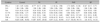
A total of 75 subjects (28 PV, 13 PF, 7 PNP, 7 BP, and 20 healthy controls) were studied. The mean±standard deviation of age was 55.28±14.10 years (range, 24~83 years) in PV patients, 51.57±18.97 (range, 15~81 years) in PF patients, 45.00±2.19 (range, 43~47 years) in PNP patients, 69.71±14.55 (range, 49~85 years) in BP patients and 24.15±3.62 (range, 17~29 years) in the healthy controls, respectively, as shown in Table 1.
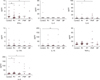
Cytokine measurements in the five groups are shown in Table 2 and Fig. 1. Cytokines which showed significant difference in the Kruskal-Wallis test was IFN-γ, IL-6, IL-10 and IL-8 (p=0.01, 0.019, 0.039 and 0.004, respectively). The median concentration of the Th1 cytokine, IFN-γ, was significantly decreased in PV and BP when compared to normal control (0.77 pg/ml in PV, 0.34 pg/ml in BP and 1.63 pg/ml in the control group; p=0.0054 and p=0.035, respectively). When comparing PV with BP, serum concentration of IFN-γ was not significantly different between the two groups (p=0.4302).
The median concentration of IL-6 was significantly higher in patients with PNP, compared to healthy control (4.92 pg/ml in PNP, 0.24 pg/ml in control; p=0.0194). IL-6 was detectable in 5 of 7 PNP sera, which all showed higher concentration than the median value of normal control sera. Higher concentrations of IL-10 were observed in the PNP group (0.86 pg/ml) compared to the normal control group (p=0.0046). IL-10 levels were higher than the median value of normal controls in all seven serum samples from the PNP group. The median concentration of IL-10 was not significantly different between the normal controls and patients with PV, PF or BP (<0.12 pg/ml in PV, PF, BP and control).
Serum levels of IL-8 were significantly higher in the PV and PNP groups than in normal controls (11.85 pg/ml in PV, 31.5 pg/ml in PNP, 8.31 pg/ml in controls; p=0.0227 and p=0.0322, respectively).
Serum levels of Th2 cytokine IL-4 were below the detection limits (<1.86 pg/ml) in all but four samples, which included one patient with BP and three healthy controls. There were no significant differences in IL-17A, the Th17 cytokine, between the five groups. TNF-α also did not differ significantly between the five groups. When comparing PV sera with PF sera, none of the cytokines showed significant difference (data not shown).
DISCUSSION
In pemphigus, it is postulated that autoreactive T cells are involved in the induction and maintenance of autoantibody production41617. It thus follows that cytokines are likely to be key players in the coordination of the cellular and humoral responses in pemphigus. The present study of serum cytokine analysis in pemphigus showed a decrease of IFN-γ in PV and BP, an increase of IL-6 and IL-10 in PNP, and an increase of IL-8 in PV and PNP when compared to healthy controls.
There have been studies reporting an imbalance of Th1/Th2 response in pemphigus, which can ultimately result in complex and severe impairment of immune function6. Once a naïve T cell differentiate to Th2 cell, specific transcription factors such as GATA-3 and c-maf are activated, which in turn activate the Th2 cytokines while down-regulating factors necessary to generate Th1 response1819. In the majority of previous studies, involvement of Th2 pathway has been demonstrated in the pathogenesis of pemphigus and as a consequence, down-regulation of Th1 pathway is to be anticipated in disease5. However, there are a considerable number of studies showing increased and decreased levels of IFN-γ, as well as studies showing no significant difference compared to control group5. The reason for the variation might potentially be due to small number of samples included in some of the studies20, and the varied timing of serum collection during the disease course21. Our result of IFN-γ was significantly decreased in PV compared to control, although it was not accompanied with the IL-4 increase. A previous study by Veldman et al.21 showed that peripheral Dsg3-reactive Th1 cells varied according to the clinical activity of PV. Sera in our study was collected in the active stage of disease, both acute and chronic, and our result suggest that the Th1 pathway may be suppressed in the active stage of PV622. In comparison of the PV sera with BP sera, which also showed decreased level of IFN-γ compared to control, there was no significant difference. In contrast to our finding, several studies reported increased level of IFN-γ in the serum and blister fluid of BP232425. On the other hand, Giomi et al.26 speculated that an early stage of BP can be characterized by an initial Th0/Th2-like response (IL-4, IL-5, low levels of IFN-γ), and a chronic Th1-skewed phase would follow. Studies in the intermediate phase would be presented by a mixed Th1/Th2 expression, this also highlighting the different cytokine profile according to disease phases. The decrease in IFN-γ in the BP group seen in the present study might represent suppression of the Th1 pathway in the active disease stage.
Even though there are some good evidences that PV is a Th2-mediated disease and several studies have demonstrated an increase in Th2 cytokines62527, we did not observe any significant differences in IL-4 between patient groups and healthy control. Although far less common, reports exist documenting reduced or no significant difference in the IL-4 level compared to control as shown in our study1520. However, it is now generally accepted that IL-4 has an important role in pemphigus pathogenesis and there is limitation in the interpretation of our result, because most samples were under the detection limit for IL-4; 74 out of 78 sera were below the detection limit (<1.86 pg/ml).
Th17 cells have been implicated in the initiation and progression of many inflammatory and autoimmune diseases such as autoimmune encephalitis, inflammatory bowel disease and psoriasis28. In pemphigus, on the other hand, there is only a limited number of data examining its role. Arakawa et al.7 found Th17 cells in skin lesions of patients with PV, but did not find a significant correlation between the Th17 cells and disease activity or anti-Dsg3 antibody titers. Giordano and Sinha5 mentioned that they had unpublished data showing an increase in IL-17A levels in PV compared to controls. However, we did not find any significant difference of IL-17A levels between the control and patient groups. Regarding the fact that Th17 cells in lesional skin neither correlated with autoantibody level nor with the clinical severity of PV, and the fact that our study showed no significant difference in serum IL-17A level, the role of Th17 cells in pemphigus still remain questionable and require further investigation.
Regulatory T cells play a crucial role in modulating peripheral tolerance and preventing autoimmunity. In pemphigus, a relative decrease in Treg function may be involved in the pathogenesis. Data reported on Treg cells and their cytokines, TGF-β and IL-10, are quite varied in pemphigus29. Cytokine studies published so far have demonstrated a lack of significant variation in TGF-β between PV and control1517. As for IL-10, a majority of published studies reported an increase in serum IL-10 in PV patients5. IL-10 does play a significant role in the Treg pathway, but it is also associated with the Th2 pathway, and some authors assumed this increase represents an activated Th2 pathway5. However in the present study, serum IL-10 values in PV, PF, and BP did not differ from control. In PNP, on the other hand, IL-10 was elevated compared to control group. IL-10 is an anti-inflammatory cytokine with a role in preventing inflammatory and autoimmune pathologies, but it is also a potent B cell stimulator that enhances activation, proliferation, and differentiation of B cells30. A recent study of PV suggested a model for the role of IL-10 in the active disease state31. It is suggested that in active PV, IL-10 favors class switch to the classic PV autoantibody subclass IgG4, and promotes B cell differentiation into antibody secreting cells which results in higher anti-Dsg3 antibody concentrations. B cells exhibiting regulatory functions (Bregs)32 are defective in active PV with reduced IL-10 secretion and inability to suppress CD4+T cell responses, which perpetuates the autoimmune reaction. In PNP, no data regarding IL-10 have been published, but our result of increased IL-10 might also reflect the autoimmune stimulating effects of IL-10 in the disease pathogenesis.
IL-6 was also significantly increased in PNP compared to healthy control, consistent with previous findings. IL-6 is known to promote differentiation and maturation of B cells, and drive immunoglobulins production. Markedly elevated serum IL-6 levels have been demonstrated in a majority of PNP patients33. Furthermore, in a subset of tumors associated with PNP, such as non-Hodgkin lymphoma, chronic lymphocytic leukemia and Castleman's disease, it has been observed that the tumor cells secrete large amounts of IL-6 in vitro34. With our result further supporting the hypothesis, IL-6 seems to contribute to the induction of autoantibodies, playing an important role in autoimmune pathology of PNP.
It may be possible to speculate that Th2 pathway is upregulated in PNP, with both IL-6 and IL-10 increased in the sera of PNP patients. IL-6 and IL-10 are also the cytokines of Th2 pathway, as well as the classic Th2 cytokine IL-4, and it is known that the Th2 cells are involved in the development of humoral immunity. As extremely limited number of cytokine studies are available in PNP, a comprehensive set of cytokines including IL-5 and IL-13 needs to be further examined to clarify our result.
Serum levels of IL-8 were significantly higher in PV and PNP compared to control. IL-8 induces chemotaxis in target cells, primarily neutrophils, and is a product of keratinocytes and dermal cells such as fibroblasts, endothelial cells and macrophages. An immunohistochemical study of pemphigus showed intensive expression of IL-8 co-localized with in vivo bound IgG in the upper epidermis where acantholysis took place35. Baroni et al.12 also demonstrated a high IL-8 value in the blister fluid of pemphigus patients, and Keskin et al.13 showed increased serum level of IL-8 compared to control. IL-8 has not been studied in PNP before, but increased serum level of IL-8 might have a role in recruitment of polymorphonuclear leukocytes to the skin lesion of PNP. However, neutrophil infiltration is not a prominent feature in PNP, and also, the small sample size of PNP sera in our study precludes a definitive conclusion. Another possible explanation for increased IL-8 in PV and PNP is that it may not be a cause but a result of the disease, i.e., an inflammatory response following the damaged epithelial barrier. It has been shown that disruptions to skin such as trauma, irritation and ultraviolet B radiation induce IL-8 in lesional skin363739.
Our study was conducted with sera collected in the active stage, and multiple blisters and erosions may trigger the release of various inflammatory cytokines including IL-8. However, it should be noted that IL-8 was not increased in PF or BP, and further detailed studies are still needed to identify the precise role of IL-8 in different pemphigus groups.
TNF-α has been widely studied in pemphigus, with majority of studies showing an increase in the serum and in the blister fluid5. It has been shown in a previous study that serum levels of TNF-α correlate with disease severity in PV15. There were also reports of anti-TNF-α drugs with certain efficacy in PV40. On the contrary, a case report of spontaneous development of PV in a psoriatic patient upon infliximab was noted41. Also, a recently published pilot study comparing etanercept versus placebo in 8 patients with PV failed to find significant therapeutic efficacy in the etanercept group42. A double-blind, placebo-controlled trial of infliximab with prednisone versus prednisone alone showed that infliximab therapy was not effective for the treatment of patients with PV43. In our study, TNF-α level in the pemphigus serum failed to show any significant difference from the control serum. While a strong increase of TNF-α was observed in the most of the previous studies, heterogeneous response of anti-TNF-α drugs in pemphigus patients imply a complex mechanism of this cytokine in the disease pathogenesis.
When comparing PV with PF, none of the cytokines we evaluated were significantly different between the two groups. Although PF and PV differ in their antigens and thus represent distinct clinical entities, concerning the pathogenic role of cytokines no major differences appear to exist between the two diseases.
The strength of our study is that we included a relatively large number of patients from a single center, and we analyzed the serum cytokine levels of PNP, which has not been studied much due to its lower incidence compared to other pemphigus subtypes. In addition, studies regarding Th17 response in pemphigus are extremely limited, and our data provides additional information in understanding the Th17 pathway in pemphigus. However, our study also has several limitations. The age and sex distributions are not homogeneous among the study group which can potentially have influenced the results. Also, the number of PNP and BP patient included in this study is relatively small and further studies with more patients will be needed in the future. Although the patients included in the study were in the active disease state with uncontrolled development of the blisters, their heterogenous treatment history including different doses of systemic corticosteroid and immunosuppressants may have influenced the serum cytokine levels.
In conclusion, our data shows decreased level of IFN-γ in PV, which may imply suppressed Th1 response in the active disease stage. A Th2 predominant response is suggested in the active stage of PNP, with elevated serum levels of IL-6 and IL-10. Increased level of proinflammatory cytokine IL-8 is observed in the sera of PV and PNP patients. Serum IL-17A levels did not differ significantly between the five groups. We think our study will be of a valuable reference data for the future studies of T cells and cytokines in pemphigus.

 XML Download