

 PDF
PDF ePub
ePub Citation
 Print
Print


INTRODUCTION
Recently, several reports about the relationship between vitamin D and many allergic diseases, including atopic dermatitis (AD), have appeared12. Some studies have indicated that vitamin D has influenced the course of immune-mediated disorders, including AD and asthma3. However, data surrounding the effect of vitamin D on the development of allergic skin diseases are conflicting. In addition, there are several debates surround the relationship between vitamin D and AD severity. Some studies demonstrated an inverse association between vitamin D levels and AD severity4567 Other studies showed no significant correlation between vitamin D levels and AD severity8910. The exact role of vitamin D in the pathogenesis of AD also has not been fully addressed.
Vitamin D, as a critical regulator of immunity, plays a role in both the innate and adaptive immune systems3. Antimicrobial defense mechanisms and epidermal barrier integrity are impaired by defective immune systems. Therefore, vitamin D deficiency might exacerbate AD via disturbed epidermal barrier function and immunologic dysregulation, with subsequent impaired defense against common allergens such as house dust mite (HDM) and infections11. Considering the functions of vitamin D, there is a possibility that vitamin D relates to HDM sensitization in AD. The effect of vitamin D on HDM sensitization in AD patients with different severity has not been established.
The aim of this study was to evaluate the correlation between serum vitamin D levels and HDM sensitization according to AD severity.
MATERIALS AND METHODS
Subjects
Data were collected from a retrospective case series of 80 patients with AD at the Department of Dermatology, Kyungpook National University Hospital, Korea, between January 2013 and September 2014. The study protocol was approved by the institutional review board of Kyungpook National University Hospital (KNUH 2015-01-002-001).
Evaluation of AD severity
Two dermatologists evaluated AD severity in all patients by means of the Rajka and Langeland score12. Grading, which may be carried out on the basis of one single consultation, permits distinction between mild, moderate, and severe AD by means of a score summation using the following parameters (each parameter had a score from 1 to 3): (1) extent (by “rule of nine”); (2) course (via history); and (3) intensity (disturbance of nightly sleep by itching)12. We classified the AD patients into two groups, either the mild to moderate group (0~7.5) or the severe group (≥8).
Laboratory determination
The Dermatophagoides farinae and D. pteronyssinus specific immunoglobulin E (IgE) levels were assayed by the immunoblot analysis (Advansure Allostation®; LG Life Sciences, Seoul, Korea) and total IgE levels were assayed by the fluorescent enzyme immunoassay (UniCAP®; Pharmacia, Stockholm, Sweden). The total IgE levels were measured up to 5,000, therefore we regarded over 5,000 as 5,000. The each HDM-specific IgE level was classified into seven quantitative classes by the following criteria: class 0, below 0.35 IU/ml; class 1, 0.35 to 0.69 IU/ml; class 2, 0.7 to 3.49 IU/ml; class 3, 3.5 to 17.49 IU/ml; class 4, 17.5 to 49.99 IU/ml; class 5, 50 to 99.99 IU/ml; and class 6, above 100 IU/ml. The patients were divided into two groups, the low sensitization group, composed of HDM-specific IgE classes 0~2, and the high sensitization group, composed of classes 3~613. Serum 25-hydroxyvitamin D3 levels were measured by an electrochemiluminescence immunoassay (Elecsys® 2010; Roche Diagnostics, Mannheim, Germany).
Assessment
To study the association between AD severity, HDM sensitization for D. farinae and D. pteronyssinus and vitamin D levels, we carried out a comparative study with the following five subsections: (1) differences in vitamin D and total IgE levels according to AD severity; (2) comparison of both HDM-specific IgE levels according to AD severity; (3) differences in vitamin D levels according to HDM sensitization; (4) relationship between vitamin D levels and HDM sensitization; and (5) relationship between vitamin D and total IgE levels.
Statistical analysis
The difference of serum vitamin D levels and serum IgE levels according to AD severity was assessed with the Mann-Whitney U-test respectively. The difference of serum vitamin D levels according to HDM sensitization was also analyzed with the Mann-Whitney U-test. Spearman's rank correlation coefficient was used to assess the relationship between vitamin D levels and HDM sensitization according to AD severity. The relationship between serum vitamin D and log transformed total IgE levels was evaluated with regression analysis and Spearman's rank correlation coefficient. A p-value of less than 0.05 was considered statistically significant. All statistical analyses were performed using SPSS Statistics ver. 17.0 (SPSS Inc., Chicago, IL, USA).
RESULTS
Patient characteristics
In total, 43 men and 37 women, mean age 19.0±11.0 years, were included in the study. We classified 35 (43.8%) and 45 (56.2%) patients into the mild to moderate AD and severe AD groups, respectively.
Differences in vitamin D and total IgE levels according to AD severity
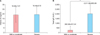
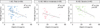
The mean serum vitamin D level was 19.29±8.03 ng/ml. There was no significant difference between serum vitamin D levels between patients with severe AD (19.46±8.10 ng/ml) and mild to moderate AD (18.98±7.97 ng/ml, p=0.72; Fig. 1A). On the other hand, the mean total serum IgE level in patients with severe AD (2,011.96±993.89 kU/L) was significantly higher than that in patients with mild to moderate AD (260.88±431.54 kU/L, p<0.05; Fig. 1B).
Comparison of HDM-specific IgE levels according to AD severity
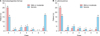
In our study, there was no significant relevance between both HDM sensitization and AD severity. However, more patients with class 6 both HDM sensitizations were found in the severe AD group (Fig. 2).
Differences in vitamin D levels according to HDM sensitization
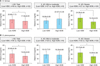
In the severe AD group, significantly lower serum vitamin D levels were found in AD patients with high D. farinae sensitization (p<0.05). However, in AD patients with mild to moderate severity, serum vitamin D levels showed no significant difference between the low and high D. farinae sensitization groups (p=0.77, Fig. 3A).
Results of D. pteronyssinus showed a similar tendency with those of D. farinae. In the severe AD group, high D. pteronyssinus sensitization group had lower serum vitamin D levels with statistical significance (p<0.05). However, there is no significant difference of vitamin D levels between the low and high D. pteronyssinus sensitization groups in mild or moderate AD patients (p=0.51, Fig. 3B).
Relationship between vitamin D levels and HDM sensitization
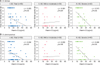
In total AD patients, vitamin D levels showed a negative correlation with D. farinae sensitization (rs=−0.283, p<0.05). In addition, there was a negative correlation between vitamin D levels and D. farinae sensitization in severe AD patients with statistical significance (rs=−0.515, p<0.05). However, no correlation was found between D. farina sensitization and serum vitamin D levels in AD patients with mild or moderate severity (rs=−0.081, p=0.64; Fig. 4A).
Results of D. pteronyssinus showed the similar findings. Vitamin D levels showed a negative correlation with D. pteronyssinus sensitization with statistical significance in total AD patients (rs=−0.254, p<0.05). In severe AD patients, serum vitamin D levels showed significantly negative correlation with D. pteronyssinus sensitization (rs=−0.484, p<0.05). However, there was no correlation between serum vitamin D levels and D. pteronyssinus sensitization in mild or moderate AD patients group (rs=−0.002, p=0.99; Fig. 4B).
Relationship between vitamin D and total IgE levels
There was a negative relationship between log transformed total IgE levels and serum vitamin D levels in total AD patients (R2=0.119, p<0.05). In addition, a negative correlation between log transformed total IgE levels and serum vitamin D levels was found in the severe AD group (R2=0.234, p<0.05). However, an association between log transformed total IgE and serum vitamin D levels was not found in AD patients with mild or moderate severity (R2=0.107, p=0.06; Fig. 5).
DISCUSSION
There are many controversies about the association between serum vitamin D levels and AD severity14. Overall, it seems that a predominance of reports points to a negative association between serum vitamin D levels and AD severity41015. However, in our study, serum vitamin D levels were not correlated with AD severity. Most AD patients have increased serum IgE levels, which correlate with disease severity16. Our results also showed a positive correlation between serum IgE levels and AD severity.
Several previous studies reported a positive association between AD severity and HDM sensitization171819. However, we were unable to replicate these findings in our study. We only found more patients with class 6 HDM sensitization in the severe AD group than in the mild or moderate groups.
A few studies examined the relationship between serum vitamin D levels and AD severity according to allergen sensitization. Akan et al.20 showed a negative correlation between AD severity and serum vitamin D levels in the group with allergic sensitization but no correlation in the group without sensitization. This study suggested that vitamin D levels in children are correlated with AD severity but only in patients with allergic sensitizations16. However, they investigated the sensitization status according to common food and aeroallergens, not specific sensitization to HDM16. Another study suggested that vitamin D deficiency increases the risk of sensitization to food allergens and that AD may be more severe in infants with vitamin D deficiency21. In our study, significantly lower vitamin D levels were found in severe AD patients with the high HDM sensitization. We thought that these results demonstrated that low serum vitamin D levels may be linked to high HDM sensitization in patients with severe AD. These results did not depend on the type of HDM, D. farinae or D. pteronyssinus.
On a molecular basis, vitamin D in the skin affects the three domains of AD pathogenesis, including the immune system, antimicrobial defense mechanisms, and epidermal barrier integrity10. Specifically, regarding its immunomodulatory effects, vitamin D influences both the innate and adaptive immune system. Vitamin D has antimicrobial effects related to macrophages and monocytes, enhancing chemotaxis and the phagocytic capabilities of innate immune cells22. In adaptive immunity, vitamin D functions in the differentiation and proliferation of T- and B-cells, leading to a shift from a proinflammatory to a more tolerogenic immune status23. Defected immune systems can influence antimicrobial defense systems and epidermal barrier integrity. Therefore, in consideration of the function of vitamin D, there is a possibility that serum vitamin D levels are associated with HDM sensitization and exaggerated immune response to HDM. Our hypothesis is that low serum vitamin D levels lead to disturbed epidermal barrier function, immunologic dysregulation, and impaired cutaneous defense mechanism in patients with an atopic background. Patients with low serum vitamin D levels have an increased risk of HDM sensitization by increased penetration of HDM through broken skin barrier. Then, high HDM sensitization may induce the aggravation of immunologic dysregulation and the development of severe AD. Findings of this study suggest that vitamin D level may affect HDM sensitization.
Regarding the relationship between serum total IgE and vitamin D levels, a previous study suggested that lower serum vitamin D levels were associated with elevated serum IgE levels624. Our study also showed that serum vitamin D levels were negatively correlated with total IgE levels in the severe AD group.
The main limitation of this study is that we did not account for clinical factors associated with serum vitamin D levels, such as individual outdoor activity and dietary habits that can affect vitamin D homeostasis. Seasonal variations in vitamin D levels were not considered. In addition, we used the Rajka and Langeland score to evaluate AD severity instead of time consuming but, more reliable scoring methods (e.g., SCORAD or EASI)25. However, recent report reintroduced the Rajka and Langeland score as a simple, useful and sensitive eczema scoring system26.
In conclusion, our results demonstrate that low vitamin D levels may be relevant to high HDM sensitization in severe AD patients. Further investigation regarding the effect of vitamin D in HDM sensitization can give new strategies for the prevention and treatment of AD. We also need the refinement and modification of large-scale studies to determine the relationship between serum vitamin D levels and HDM sensitization in AD.
 XML Download