

 PDF
PDF ePub
ePub Citation
 Print
Print


INTRODUCTION
Research on dental color and studies of tooth discoloration are important topics, with significant practical implications.123 Dental clinicians and researchers in the field of esthetic dentistry are challenged to understand the optical properties of teeth and their application in everyday practice. Dental color evaluation is a critical component of esthetic dentistry. It requires knowledge on the dimensions of tooth color and on the available tools used for their assessment, as well as clinical experience and technical skills to transfer these parameters towards a natural outcome of dental restorations.
Every direct or indirect restoration is evaluated in terms of color, shape, and texture.4 However, color appears to be the most important factor for most patients.5
An adequate definition of color was published in 2001 by the Commission Internationale de l'Eclairage (C.I.E.), as being a “characteristic of the visual perception that can be described by the attributes of hue, value, and chroma”. Used almost exclusively in color research, the CIE L*a*b* system describes color as being the product of blending three color coordinates: L* (Luminance or lightness value), a* (color coordinate on the red-green axis), and b* (color coordinate on the yellow-blue axis). By specifying these three numerical values, the CIE L*a*b* system is able to locate the color of an object in a three-dimensional (3D) color space.6
Hue is the angular component of a polar representation of the CIE L*a*b* color space, in which chroma is its radial component. The hue angle (h), defined as h = artg (b*/a*) and measured in degrees, is the dimension of color related to the perceived light wavelengths. Teeth have been described as having their hue in the range of yellow-brown, with different degrees of saturation.
Value (L*) is thought to be the most important dimension of color in dentistry.7 A color's L* is defined by the amount of black and white within the scale, which is related to lightness/darkness, whilst chroma (C*) represents the degree of saturation of a color.68
In previous studies,910 variations of color coordinates have been reported for human teeth in the range of L* = 60 − 95 (where 0 = black and 100 = white), b* = 8 − 25 (positive values designate colors towards yellow, negative values designate colors towards blue), and a* = −2 to + 10 (negative values designate colors towards green, positive values designate colors towards red). The CIE L*a*b* color space is also used to characterize changes in tooth color. For example, when teeth are whitened with hydrogen peroxide, they become brighter (L* increases), less red (a* increases in positive values), and less yellow (b* increases in positive values).111213
The evaluation of tooth shades may be a very difficult task for every clinician.14 It can be achieved either by visual comparison of the target tooth with tabs from commercial shade guides, or by measuring the color coordinates with devices based on colorimetry, spectrophotometry, spectroradiometry, or digital imaging. Instrumental shade matching techniques have been introduced and advocated to reduce the subjectivity of visual shade selection.8 Compared to conventional visual shade assessment, spectrophotometric analyses were determined to be more reproducible.815
Pathology consisting in the loss of tooth vitality has various and complex etiologies and, in addition to other consequences, it can also influence dental color. Changes in the initial hue, saturation, and lightness value, which appear after the loss of tooth vitality, immediately or in time, are also influenced by the endodontic treatment protocol, materials and irrigants used, and by the quality of the coronal reconstruction.23 Quite often, the first clue suggesting the need for further investigation is dental color, which is perceived as being different, compared to a corresponding vital tooth.
Numerous studies have focused on the evaluation of dental color parameters, either in vivo or on extracted teeth, with the aim to define a natural tooth color space, within a population.91617 The measurements involved the whole visible tooth surface, or different dental areas1819 and they were interpreted in respect to age, sex, or skin color,152021 as well as the position of the tooth in the dental arch.2223
However, although these studies concluded on the values of dental color parameters, no attempts have been made to differentiate tooth colors that characterize vital teeth, in comparison to the color parameters of non-vital ones. Moreover, although the modifications in dental color due to the loss of tooth vitality are well known, less information is available regarding the degree of tooth discoloration and the difference between the color of non-vital teeth and that of their corresponding vital teeth.
The aim of this study was to define a tooth color-space for non-vital teeth and to compare it with the color-space of their corresponding vital teeth. The null hypothesis was that no significant differences may be found between the values of L*, a*, b*, C*, h in non-vital teeth, compared to the corresponding values in matched vital teeth of the same patients.
Go to : 
MATERIALS AND METHODS
The study targeted patients with non-vital teeth from Cluj-Napoca, Romania. It included 218 consecutive patients with non-vital teeth, who addressed the Department of Propaedeutics and Esthetic Dentistry at the Faculty of Dentistry of the Iuliu Hatieganu University of Medicine and Pharmacy, in Cluj-Napoca, Romania, as well as a private dental office from Cluj-Napoca, Romania.
The studied sample included 359 non-vital and 286 vital teeth from these 218 patients (130 female, F and 88 male, M), with the age range between 17 and 70.
All participants were provided with information on the study protocol and signed an informed consent, approved by the Research Ethics Committee of the Iuliu Hatieganu University of Medicine and Pharmacy, Cluj-Napoca.
The color parameters of the 359 non-vital teeth, 91 anterior teeth (55 central, and 36 lateral incisors), 24 canines, and 244 posterior teeth (119 premolars, and 125 molars) were measured with a clinical spectrophotometer (Vita Easyshade Advance, Vita Zahnfabrik). Lightness (L*), chromatic parameters (a*, b*), chroma (C*), hue angle (h) and the closest Vita Shade in both Classical and 3D Master codifications were recorded.
The contact probe tip with the protective shield was held at 90° to the tooth surface, in its middle third, the area that best illustrates tooth shade.9 A custom made acrylic positioning device was used to adapt the probe tip on the tooth surface, in order to measure similar tooth areas and to avoid variations of the probe tip angulation. Three readings were recorded for each measurement in Single Tooth mode. An average of the three measurements was used for further analysis, in every case.
For every patient, the same color parameters were also recorded for a vital reference tooth. Therefore, the same measurement protocol has been applied to a total of 286 vital reference teeth, 86 anterior teeth (56 central incisors, 30 lateral incisors, 26 canines) and 174 posterior teeth (110 premolars and 64 molars). Whenever possible, the matching contralateral vital tooth was measured as reference, in order to provide the best possible matching for comparing dental color parameters of vital and non-vital teeth. In patients with several non-vital measured teeth, the same vital reference tooth was used as reference.
The inclusion criteria of the measured teeth were: intact crowns or with limited composite resin reconstructions, not affecting the buccal surface of the tooth, thereby allowing a correct positioning of the measuring device. Teeth with extensive fillings or prosthetic reconstructions of the buccal surface, teeth with untreated caries and temporary fillings were excluded from both the vital and the non-vital teeth group. Participants who reported tooth bleaching within the previous 6 months were also excluded. If the corresponding tooth was absent or non-vital or did not meet the inclusion criteria due to extensive fillings, crowns, or veneers, the closest sound vital tooth was measured and used as reference.
The measurements were performed by the same operator, with the same spectrophotometer, using the standardized protocol for color evaluation described above. The characteristics of the artificial illuminants were identical in both dental offices (ceiling fluorescent tube lighting OSRAM LumiluxDeluxe 36W/965 5500K).
The measured teeth were grouped in: Group 1 = vital incisors (VI) - central and lateral incisors, Group 2 = vital canines (VC), Group 3 = vital posterior teeth (VP) - premolars and molars, Group 4 = non-vital incisors (NVI), Group 5 = nonvital canines (NVC), Group 6 = non-vital posterior teeth (NVP).
Color differences between vital and non-vital teeth were calculated using the CIEDE2000 color-difference formula (ΔE00), which includes lightness, chroma, and hue weighting functions, as well as a scaling factor for a* and a rotation function (RT) between chroma and hue differences, in order to improve the formula performance for grey and blue colors:24
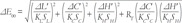
Since the lighting conditions in this study were standardized, the parametric correction factors, KL, KC, and KH were set at values of 1.
All color parameters (L*, a*, b*, C*, h) have been compared between vital and non-vital teeth, using Wilcoxon tests for paired samples. A Bonferroni correction from α = 0.05 down to α = 0.01 has been used as a statistical significance threshold level, in order to account for these multiple comparisons.
For the pairs of vital-nonvital contralateral teeth (for instance, vital maxillary canine in one quadrant vs non-vital maxillary canine in the other quadrant), the color difference, ΔE, was calculated according to the CIEDE2000 color difference formula for each of the three groups of teeth (VI vs NVI, VC vs NVC, VP vs NVP). These computed color differences were then compared among incisors, canines, and posteriors, as well as among patients from different age groups, using a Kruskal-Wallis rank sum test, followed by U Mann-Whitney rank sum tests with continuity correction and a Bonferroni correction of the statistical significance threshold level down to α = 0.017, for post-hoc testing.
Only the 332 matching pairs of vital and non-vital teeth were included in the ΔE comparisons. The remaining 27 pairs of teeth, belonging to different groups, due to unavailable same-group vital reference teeth (for instance, lateral non-vital tooth vs frontal reference tooth), were only included in the overall comparison of color parameters (L*, a*, b*, C*, h) between vital and non-vital teeth.
Data analysis has been performed using Microsoft Excel 2010 (Microsoft, Redmond, WA, USA), R 3.1.1. (R Foundation for Statistical Computing, Vienna, Austria) and OriginPro 8.0724 (OriginLab, Northampton, MA, USA).
Go to :
RESULTS
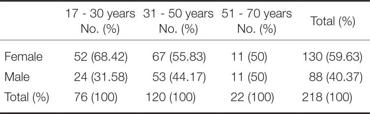
Most included patients (59.63%) were female and belonged to the middle aged group (55.04%). Patient distribution based on age and sex is presented in Table 1.
In non-vital teeth, L* values were lower, compared to vital teeth. C* values exhibited higher values, while the hue (h) interval was wider for non-vital teeth compared to vital ones. The color coordinates of non-vital teeth varied as follows: lightness L*: 52.83 – 92.93, C*: 8.23 – 58.90, h: 51.20 – 101.53, a*: −2.53 – 24.80, b*: 8.10 – 53.43. For the reference vital teeth, the ranges of color parameters were: L*: 60.90 – 97.16, C*: 8.43 – 39.23, h: 75.30 – 101.13, a*: −2.36 – 9.60, b*: 8.36 – 39.23.
Table 2 presents the main descriptive statistics of color coordinates for vital and non-vital teeth.
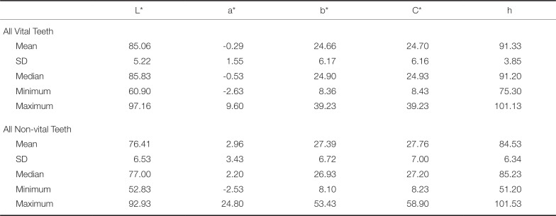
Table 2
Descriptive statistics of color parameters for vital and non-vital teeth in the studied sample
![]()
All color parameters (L*, a*, b*, C*, h) differed significantly between vital and non-vital teeth (P < .0001 - Wilcoxon test for paired samples). The chromatic color coordinates (a* and b*) displayed a wider range for non-vital teeth compared to vital teeth, being especially grouped towards positive values.
The CIE L*, a*, b* color space of vital versus non-vital teeth is illustrated in Fig. 1. The lightness was concentrated in a low-value area for the non-vital teeth and in a higher-value area for vital teeth. The corresponding grouping pattern of a* and b* chromatic parameters in vital versus non-vital teeth is also presented in Fig. 2. An increased tendency towards positive values on the a* and b* axes of non-vital teeth has been observed, indicating redder and yellower non-vital teeth in comparison with vital ones.
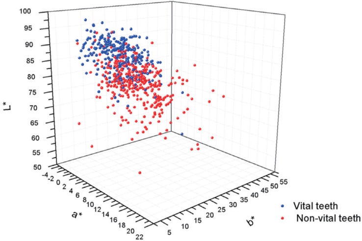
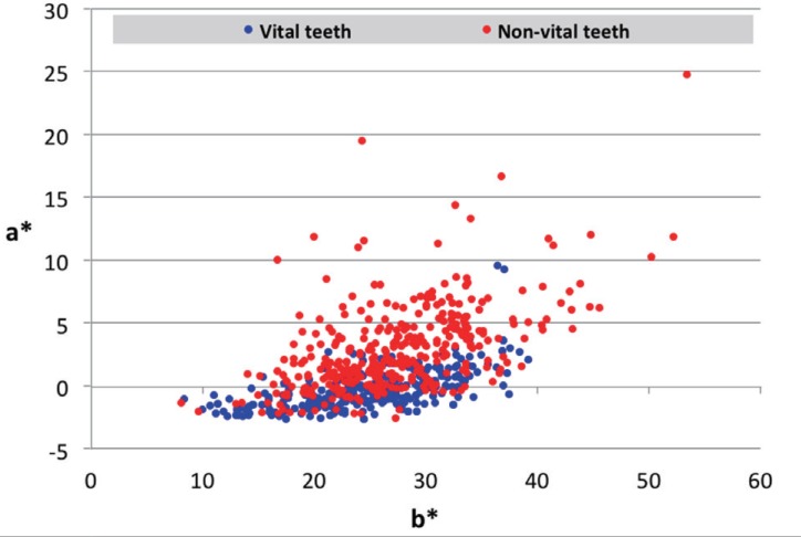
Table 3 contains the color coordinates of each group of teeth (Vital incisors VI, Vital canines VC, Vital posterior teeth VP, Non-Vital incisors NVI, Non-Vital canines NVC, Non-Vital posterior teeth NVP). The highest lightness and lowest chroma were found for VI, while the lowest lightness and most increased chroma were found for NVP. The highest a* and b* values were found in NVP and the lowest in VI.
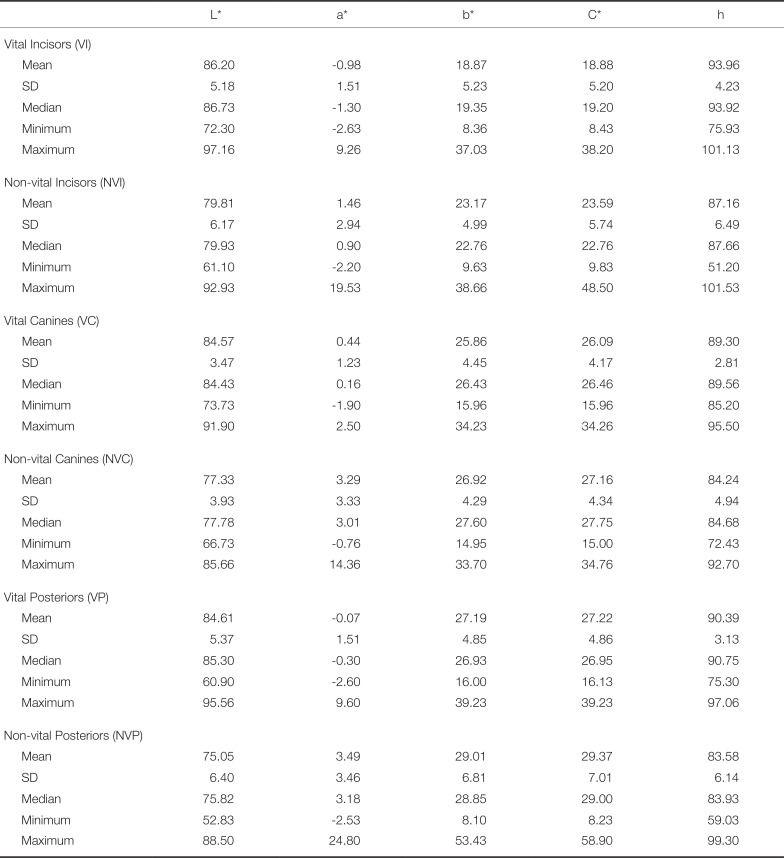
Table 3
Descriptive statistics of color parameters in the vital and non-vital teeth groups
![]()
The three most frequent values found using the Vita Classical shade guide were B3 (25.63%), A4 (25.07%), and A3,5 (12.53%), for non-vital teeth, and B3 (25.07%), A3 (22.01%), and A1 (15.04%), for vital teeth. Using the 3D Master shade guide, the three most frequently recorded shades for non-vital teeth were 2M3 (18.66%), 3M3 (156%), and 2R2.5 (11.98%), while vital teeth were most frequently recorded as having 2M3 (23.68%), 2M2 (22.01%), and 1M2 (14.76%) shades. Table 4 and Table 5 illustrate the distribution of color shade in vital and non-vital teeth.
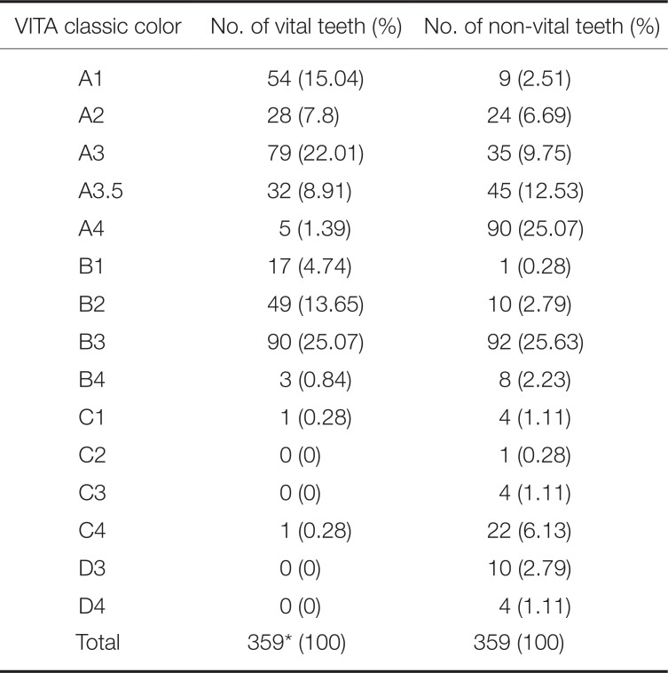
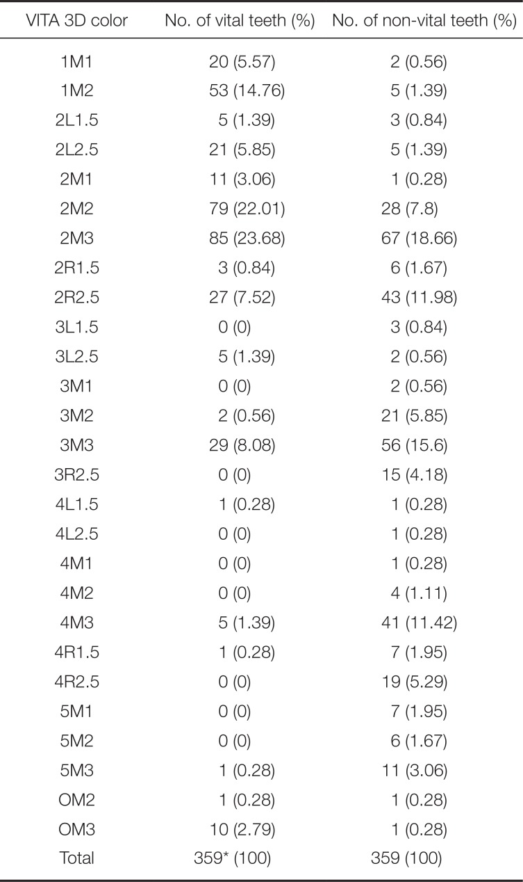
Table 4
Distribution of measured Vita Classic shades in vital and non-vital teeth
![]()
Table 5
Distribution of measured Vita 3D Master shades in vital and non-vital teeth
![]()
This study found color differences between vital and non-vital teeth (CIEDE2000 color difference formula) ranging between ΔE00 = 0.42 - 27.20, with a median ΔE00 = 7.10. Three out of the 332 ΔE values between matching pairs of vital and non-vital teeth fell under the perceptibility threshold of ΔE00 = 0.8,25 and 11 fell under the acceptability threshold of ΔE00 = 1.8.25 Among the 27 non-matching pairs of vital and non-vital teeth, one more ΔE value fell below the acceptability threshold of ΔE00 = 1.8. Therefore, from a total of 359 ΔE00 values, 12 were in the acceptable and 3 were in the non-perceptible range.
Fig. 3 illustrates the frequency distribution of ΔE00 color differences between the 332 matching pairs of vital and non-vital teeth.
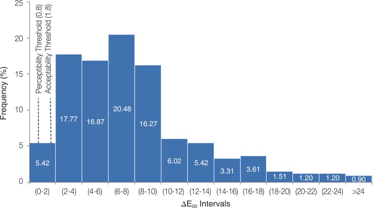
Delta E00 between vital and non-vital teeth differed significantly (P = .013 - Kruskal-Wallis test) between Incisors (median ΔE00 = 6.015), Canines (median ΔE00 = 6.374) and Posterior teeth (median ΔE00 = 7.452).
In post-hoc testing, the color difference ΔE00 of Incisors (VI vs NVI) differed significantly from the ΔE00 of Posteriors (VP vs NVP) (P = .010 - U Mann-Whitney test), while ΔE00 of Canines vs. ΔE00 of Posterior teeth did not reach statistical significance (P = .067 - U Mann-Whitney test). Nevertheless, there was a clear difference in respect of ΔE00 between anterior teeth (Incisors & Canines) and posterior teeth (P = .0035 - U Mann-Whitney test).
No significant differences in respect to ΔE00 between vital and non-vital teeth were found, neither among young, middle-aged, and elderly patients (P = .286 - Kruskal-Wallis test) nor between sexes (P = .217 - U Mann-Whitney test).
Go to :
DISCUSSION
The null hypotheses of the study was rejected, after finding highly significant differences among all color coordinates of investigated vital versus non-vital teeth. The color space of vital teeth was partially within the color space of non-vital teeth, but the color space of non-vital teeth was wider.
The a* and b* coordinates of non-vital teeth extended farther on the positive axis (red and yellow) and less on the negative axis (green and blue), confirming non vital teeth to be more chromatic than vital teeth (redder and yellower). Another finding was that lightness (L*) of non-vital teeth exhibited smaller values compared to vital teeth, while their chroma (C*) shifted towards values of higher color saturation. As previously suggested in literature,3 such findings can be explained by endodontic pathology, treatment protocol, or dental materials. In these cases, chromogen agents, originating from the dental pulp or endodontic materials that impregnate the dental structure can be responsible for changes in dental color. However, the evaluation of dental color in saturated, darker teeth is more difficult, compared to lighter teeth.16
In the present study, the Vita Classic B3 shade was found to be most frequent in both non-vital and vital groups. This can be explained by the higher prevalence of posterior non-vital teeth in the studied sample, along with their matching vital teeth (a total of 418 posterior versus only 177 frontal teeth). These results were in agreement with other published literature, in which B3 has been reported as being the most frequent shade of posterior vital teeth.22
For both vital and non-vital teeth, 2M3 was the most frequent Vita 3D Master shade. This was in agreement with studies that evaluated both frontal and posterior vital teeth.22 The results of the current study differed from those of other researchers, who reported 2M1 (Yuan et al.16), A3 and 3M1 (Gómez et al.15), A3 and 1M2 (Elamin et al.20) and 2R2.5 (Rodrigues et al.21) as the most frequently measured shade; however, the evaluated teeth in these studies were only vital central incisors.
In 2015, Paravina et al.25 reported a clinical perceptibility threshold (50:50%) of ΔE00 = 0.8 and an acceptability threshold of ΔE00 = 1.8. Most ΔE00 color differences between vital and non-vital teeth found in the current study (N = 347) exceeded the acceptability threshold24 of ΔE00 = 1.8. Only 12 of all 359 ΔE00 values fell below the acceptability threshold25 of ΔE00 = 1.8, including three values that also fell below the perceptibility threshold25 of ΔE00 = 0.8. This confirmed the need to improve the color of the non-vital teeth, to match the vital correspondent ones, as a clinically important outcome.
No difference based on age-group or gender was found in our study regarding the color difference between vital and non-vital teeth. This suggests that the progression of dental color in non-vital teeth is probably not influenced by general factors such as age and gender.
Vita Easyshade is a widespread shade measuring device, extremely accurate both for in vivo and in vitro studies.2627 A limitation of this device may derive from the edge loss phenomenon, due to the incongruence between the two surfaces set in contact (tooth and probe). To avoid probe angulation, a custom made acrylic tray was used for every measurement. The middle third of the tooth was chosen as measurement site because it is generally flatter, and it is the most representative for tooth color.1621 The incisal area is too translucent and it allows light to be transmitted, rather than reflected to the spectrophotometer.1628
Another limitation of the current study may have been the relatively higher prevalence of posterior non-vital teeth in the studied sample, compared to anterior ones (244 posteriors, 91 incisors, and 24 canines, non-vital teeth). This may have been responsible for the concentration of measured shades in the Vita Classic B3 shade, for both vital and non-vital teeth, as the tooth color of posterior teeth has decreased lightness and increased saturation.23 Nevertheless, given that the current study has included consecutive patients with non-vital teeth who addressed both a private dental office and a University clinic of the same city, this frequency distribution may in fact represent a close estimate of the actual prevalence distribution of non-vital teeth in the target population of the current study.
Non-vital teeth that met the inclusion and exclusion criteria of this study have been analyzed in order to define their color space. Moreover, the same vital reference tooth was used more than once in patients who had more than one non-vital tooth, whenever the other matching vital tooth did not meet the defined inclusion criteria.
The current study defined a color space for non-vital teeth that only partially overlapped on the space color of the matching vital reference teeth. Previous studies91819 have also tried to demonstrate that currently available shade guides are insufficient to cover the natural tooth color space. However, these comparisons have been performed either between available shade guides and vital tooth measurements, or, without accounting for the vital or non-vital status of the studied teeth.
Tooth color of vital teeth has been clinically measured by other researchers in order to establish a tooth color space. Different devices have been used (colorimeters, spectrophotometers, spectroradiometers) and large variations of L*, a* and b* have been reported. Odioso et al.29 reported mean values of L* = 69.3, a* = 5.4, and b*= 18.7. Hasegawa et al.18 reported mean values of 73.1 for L*, 3.4 for a*, and 16.4 for b*. Zhu et al.9 conducted a study on vital frontal teeth, both maxillary and mandibular, and reported a large range for L* (21.89 – 83.75), and intervals between −8.07 – 9.21 for a* and −6.53 – 59.89 for b*. Tooth color was measured with a dental spectrophotometer in all three studies. For vital central incisors, Gozalo-Diaz et al.30 reported that L* ranged between 38.0 and 89.5, a* between 0.3 – 12.2 and b* between 5.7 – 35.7. In their study, the device for color measurements was a colorimeter. Results can vary a lot when using different devices for tooth color measurements due to their different sensitivities, as exemplified by the study of Cho et al.31
Compared to previously published findings, a larger range of color coordinates (L*, a*, b*) was found in the current study. This fact can be explained by the large number of both vital and non-vital, incisors, canines and posterior teeth included in this study. When considered separately, a very high mean value of L* values was found in the current study for vital teeth (85.6) and a very large range for chromatic parameters a* and b* in non-vital teeth: −2.53 – 24.80 for a* and 8.10 – 53.43 for b*. These findings suggest that non vital teeth have a broader color space compared to vital teeth. Further research may be necessary to verify these findings and to expand the currently accepted color range of natural teeth.
Defining a tooth color space is a very challenging theme for dental researchers. Direct and indirect restoration of non-vital teeth must be considered in terms of both esthetics and biomechanical appearance. Regardless of the great variety of existing dental shades, color of non-vital teeth has a great impact on the final restauration. When it comes to treatment needs, if the color difference between vital and non-vital teeth exceeds the perceptibility and acceptability thresholds, a requirement for corrective therapy can be expected.
Go to :
CONCLUSION
Within the limitations of the present study, we can conclude that non-vital teeth have a wider color space than vital ones.
Differences have been found between the spectra covered by color coordinates of non-vital teeth compared to the matching vital teeth used as reference. Non-vital teeth appear to be darker (decreased lightness) and more saturated (increased chroma), and have an increased hue interval, compared to their vital counterparts. There was an increased tendency towards positive values on both the a* and the b* axes, suggesting more reddish and yellowish non-vital teeth compared to vital ones. The color differences between vital and non-vital teeth depended on tooth group (more important in posterior teeth) but not on patient age. In most investigated pairs, ΔE00 between vital and non-vital teeth exceeded the perceptibility and acceptability thresholds.
Go to :
 XML Download