Abstract
Purpose
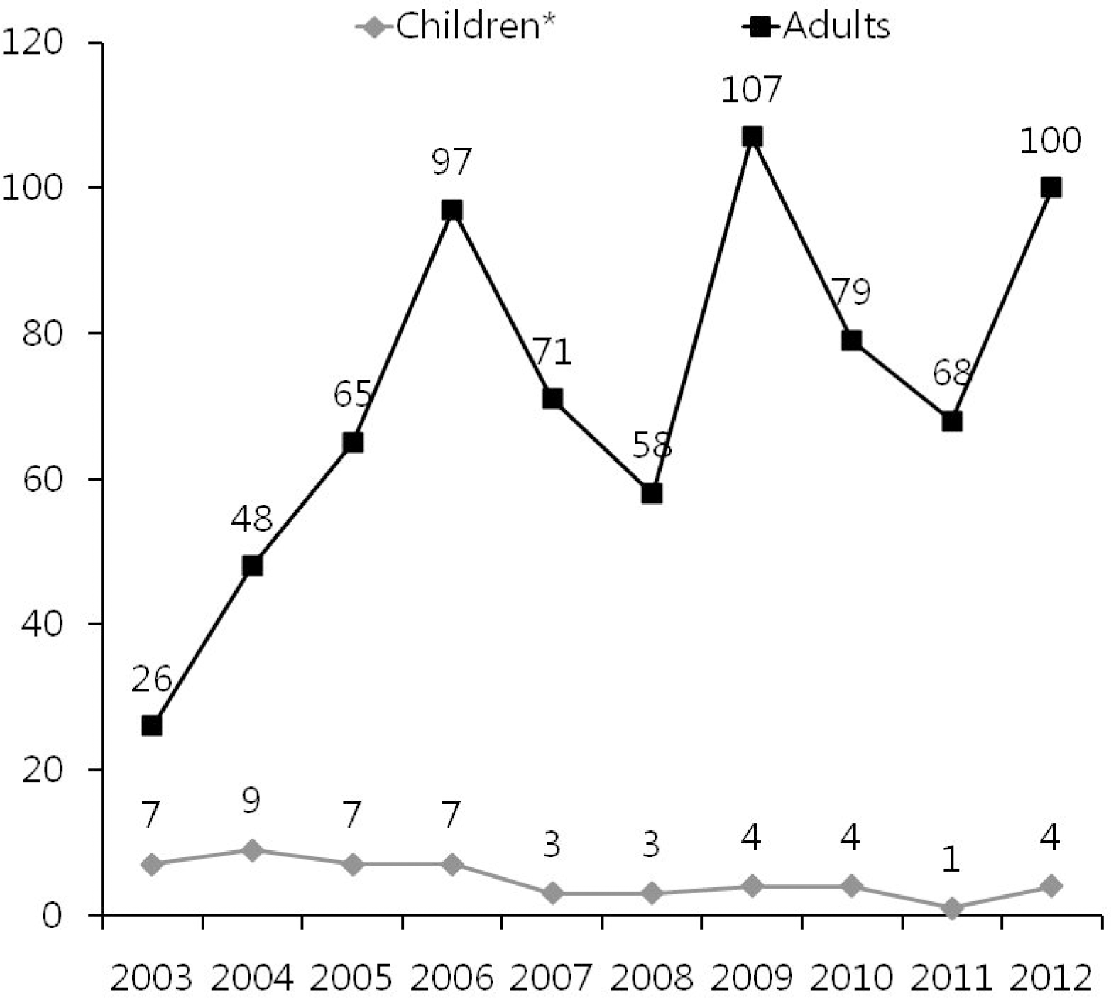
We compared the clinical manifestations of patients with tsutsugamushi disease between children and adults. Methods: From January 2003 to December 2012, 768 patients diagnosed with tsutsugamushi disease were retrospectively reviewed, and the clinical characteristics, laboratory findings, and complications were compared between children and adults. Results: No patterns of annual increases in the number of patients were noted in both children and adults. The higher incidences occurred in October and November respectively. By gender, male outnumbered female in children, but the opposite trend was seen in adults. By residential area, the urban distribution of children was higher than that of adults. Rashes (P=0.001) and eschar (P=0.004) were more common in children, while myalgia was more common in adults. Child-ren had a high prevalence of anemia (P=0.041), and low incidence rates of thrombocytopenia, abnormal liver and renal function. Children yielded better results in the duration of their hospital stay and the incidence of complications (P˂0.001). A comparison of the therapeutic effects of doxycycline and macrolide antibiotics, which was performed only on the children, did not reveal any significant differences. Conclusion: Compared to adults, children had higher incidence rates of male patients and more often suffered from rashes and eschar. Children yielded better results in the laboratory findings and duration of the hospital stay and complications. Therefore, when children are suspected to have tsutsugamushi disease, especially during its peak occurrence period, detailed physical examination and serological test should be performed to ensure a prompt diagnosis, and the use of macrolide antibiotics, which have fewer side effects, is expected to yield the same therapeutic effects.
References
1. Jang JG, Park PG, Lee HS, Maeng JH, Kim HS, Lee SC, et al. The study of 46 cases of tsutsugamushi disease in Young-Dong region in Gang-Won-Do. J Infect Chemother. 2003; 25:138–44.
2. Lee HG, Min SK, Kong SJ, Lee SJ, Song HH, Yoon JW, et al. Clinical features of tsutsugamushi disease in Chuncheon. Korean J Med. 2005; 69:190–6.
3. Kim KJ, Cho NS, Cho SH. Related clinical finding result on complication of tsutsugamushi patients. J Korean Soc Emerg Med. 2001; 12:268–76.
4. Chang WH. Scrub typhus. J Korean Med Assoc. 1994; 37:1400–7.
5. Hong CE. Textbook of pediatrics. 10th ed.Reston: MiraeN Co.;2012. p. 434–6.
6. Park HJ, Lee KY. Roxithromycin treatment of tsutsugamushi disease (scrub typhus) in children. J Korean Pediatr Soc. 2003; 46:710–3.
7. Park BK, Kim SH, Oh YK, Yoon HS, Uhm MK, Yoo HW, et al. Clinical features and serial changes in the indirect immunofluorescent antibody titers by the duration of illness in 28 children with scrub typhus. Korean J Infect Dis. 1993; 25:109–23.
8. Yi KS, Chong YS, Chun CH, Tsunehisa Suto. Importance of finding eschar in the early diagnosis of tsutsugamushi disease. J Korean Med Assoc. 1987; 30:1009–16.
9. Song JY, Han JW, Hwang SS, Lee KY, Lee KS. A clinical study of tsutsugamushi disease in children. J Korean Pediatr Soc. 1995; 38:641–8.
10. Ju HY, Lee JS, Kim JH, Yoo HJ, Kim CS. A clinical study of tsutsugamushi fever in children during 1997–2000 in the western Kyungnam Province. Korean J Pediatr Infect Dis. 2001; 8:213–21.

11. Kim S, Jung EM, Moon KH, Yoe SY, Eum SJ, Lee JH, et al. Clarithromycin therapy for scrub typhus. Korean J Pediatr Infect Dis. 2002; 9:175–81.
12. Lee JS, Ahn CR, Kim YK, Lee MH. Thirteen cases of rickettsial infection including nine cases of tsutsugamushi disease first confirmed in Korea. J Korean Med Assoc. 1986; 29:430–8.
13. Chang WH, Kang JS. Isolation of Rickettsia tsutsugamushi from Korean patients. J Korean Med Assoc. 1987; 30:999–1008.
14. Chang WH, Choi MS, Park KH, Lee WK, Kim SY, Choi IH, et al. Seroepidemiological survey of tsutsugamushi disease in Korea, 1987 and 1988. J Korean Soc Microbiol. 1989; 24:185–95.
15. Chang WH, Kim IS, Choi MS, Kee SH, Han MJ, Seung SR, et al. Seroepidemiological survey of scrub typhus in Korea, 1991. J Korean Soc Microbiol. 1992; 27:435–42.
16. Hame TK, Kim SC, Bae CW, Choi YM, Ahn CI. A case of tsutsugamushi disease. J Korean Pediatr Soc. 1988; 31:1048–53.
17. Jee ES, Chung HL, Lee HS, Moon HR. Two cases of tsutsugamushi disease in Children. J Korean Pediatr Soc. 1988; 31:1509–15.
18. Lee DG, Kim SH, Han BK, Lee KH, Hwang CH, Cho MK. The clinical study of 10 cases of tsutsugamushi fever. J Korean Pediatr Soc. 1994; 37:689–94.
19. Lee JY, Lee SB, Do BS. Clinical features of tsutsugamushi disease in the emergency department. J Korean Soc Emerg Med. 2009; 20:569–76.
20. Hong JH, Oho JH, Kim JH, Kho DK. A case of acute renal failure associated with tsutsugamushi disease. J Korean Pediatr Soc. 2001; 44:464–8.
21. Jang KM, Kang MH, Yang YS, Hwang HG, Lee KP, Lee JS, et al. The twenty cases of serologically confirmed tsutsugamushi disease. J Korean Med Assoc. 1987; 30:638–46.
22. Lee BG, Park TH, Cho SC, Lee DY, Kim JS. 3 cases of tsutsugamushi disease with meningitis in children. Korean J Infect Dis. 1993; 25:183–7.
23. Lee HW, Chu YK, Choi KY, Kim YS, Kim MJ, Park SC. Serologic diagnosis, epidemiology and clinical features of scrub typhus in Korea in 1985. Korean J Infect Dis. 1988; 20:83–92.
24. Kim YK, Kim JM, Kim E, Chung DK, Ham YH, Hong CS. A clinical study of tsutsugamushi disease occurred in Seoul and Kyungki do in autumn of 1987. Korean J Infect Dis. 1988; 20:93–103.
25. Lee JS, Kang JH, Cho BK, Yu BY. Factors affecting disease duration in patients with tsutsugamushi disease. J Korean Acad Fam Med. 2007; 28:774–81.
26. Kim YO, Jeon HK, Cho SG, Yoon SA, Son HS, Oh SH, et al. The role of hypoalbuminemia as a marker of the severity of disease in patients with tsutsugamushi disease. Korean J Med. 2000; 59:516–21.
27. Kim EJ, Lee CY, Oh YG, Yun HS, Kim JD. Four cases of scrub typhus treated with azithromycin in children. J Korean Pediatr Soc. 2003; 46:188–91.
28. The Korean society of infectious diseases. Antibiotic guideline. 3rd ed.Reston: MIP Co.;2008. ;110.
Fig. 1.
Comparison of number of patients with tsutsu-gamushi disease between children and adults during 2003–2012.∗ Children defines under 15-year-old.

Table 1.
Comparison of Distribution of Patients with Tsutsugamushi Disease between Children and Adults by Months
| Months | No. of patients (%) | |
|---|---|---|
| Children∗ | Adults | |
| January | 0 (0.0) | 11 (1.5) |
| February | 1 (2.0) | 3 (0.4) |
| March | 0 (0.0) | 2 (0.3) |
| April | 0 (0.0) | 5 (0.7) |
| May | 0 (0.0) | 4 (0.6) |
| June | 0 (0.0) | 5 (0.7) |
| July | 0 (0.0) | 2 (0.3) |
| August | 0 (0.0) | 9 (1.3) |
| September | 1 (2.0) | 12 (1.7) |
| October | 19 (38.8) | 350 (48.7) |
| November | 28 (57.1) | 296 (41.2) |
| December | 0 (0.0) | 20 (2.8) |
| Total | 49 (100.0) | 719 (100.0) |
Table 2.
Comparison of Distribution of Patients with Tsutsugamushi Disease between Children and Adults by Sex
| Group | Sex | Ratio | P-value | |
|---|---|---|---|---|
| Male (%) | Female (%) | |||
| Children∗ | 26 (53.1) | 23 (46.9) | 1.13:1 | 0.029 |
| Adults | 269 (37.4) | 450 (62.6) | 0.60:1 | |
Table 3.
Distribution of Patients with Tsutsugamush Disease in Children According to Age and Sex
| Age (years) | Sex | No. of patients (%) | |
|---|---|---|---|
| Male (n=295) | Female (n=473) | ||
| <1 | 2 | 0 | 2 (0.3) |
| 1–5 | 13 | 12 | 25 (3.3) |
| 6–10 | 9 | 9 | 18 (2.3) |
| 11–14 | 2 | 2 | 4 (0.5) |
| ≥15 | 269 | 450 | 719 (93.6) |
Table 4.
Comparison of Clinical Findings of Patients with Tsutsugamushi Disease between Children and Adults
Table 5.
Comparison of Location of Eschar in Patients with Tsutsugamushi Disease between Children and Adults
| Location | No. of eschar (%) | |
|---|---|---|
| Children∗ | Adults | |
| Trunk | 11 (22.4) | 228 (38.0) |
| Extremity | 11 (22.4) | 166 (27.8) |
| Head and neck | 9 (18.4) | 43 (7.2) |
| Inguinal area | 5 (10.2) | 55 (9.2) |
| Hip | 5 (10.2) | 16 (2.7) |
| Axillary area | 4 (8.2) | 62 (10.3) |
| Genitalia | 3 (6.1) | 19 (3.2) |
| Unknown site | 1 (2.0) | 10 (1.6) |
| Total | 49 (100.0) | 599 (100.0) |
Table 6.
Comparison of Laboratory Findings of Patients with Tsutsugamushi Disease between Children and Adults
| Laboratory findings | No. of patients (%) | P-value | |
|---|---|---|---|
| Children∗ | Adults | ||
| Hematologic | |||
| Anemia (<11 g/dL) | 13 (26.5) | 100 (15.4) | 0.041 |
| Leukocytosis (≥10,000/μ L) | 5 (10.2) | 103 (15.8) | 0.292 |
| Leukopenia (<5,000/μ L) | 19 (38.8) | 205 (31.5) | 0.295 |
| Thrombocytopenia (<150,000/μ L) | 20 (40.8) | 395 (60.7) | 0.006 |
| Liver function | |||
| Elevated AST (≥40 U/L) | 42 (85.7) | 546 (81.7) | 0.484 |
| Elevated ALT (≥40 U/L) | 21 (42.9) | 434 (65.1) | 0.002 |
| Hypoalbuminemia (<3.0 g/dL) | 0 (0.0) | 92 (14.2) | 0.005 |
| Renal function | |||
| Elevated BUN (≥20 mg/dL) | 0 (0.0) | 188 (29.0) | 0.001 |
| Elevated Cr (≥1.2 mg/dL) | 0 (0.0) | 161 (24.8) | 0.001 |
| Urine analysis | |||
| Proteinuria (≥2+, 100 mg/dL) | 0 (0.0) | 104 (16.5) | 0.002 |
| Hematuria (≥2+, 50/μ L) | 0 (0.0) | 116 (18.4) | 0.001 |
| Chest X-ray | |||
| Pneumonia | 1 (2.0) | 38 (5.8) | 0.275 |
| Pleural effusion | 1 (2.0) | 27 (4.1) | 0.483 |
| Others† | 0 (0.0) | 14 (2.1) | 0.305 |
Table 7.
Comparison of Seropositive Cases of Patients with Tsutsugamushi Disease between Children and Adults
| Group | No. of seropositive∗cases (%) | P-value |
|---|---|---|
| Children† (n=39) | 23 (59.0) | 0.210 |
| Adults (n=650) | 447 (68.8) |
Table 8.
Comparison of Duration of Hospitalization of Patients with Tsutsugamushi Disease between Children and Adults
| Duration of hospitalization | Group | P-value | |
|---|---|---|---|
| Children† (n=49) | Adults (n=719) | ||
| Mean (day†) | 5.61±1.39 | 8.66±8.20 | <0.001 |
| <1 week (%†) | 38 (77.6) | 280 (38.9) | |
| ≥1 week (%†) | 11 (22.4) | 439 (61.1) | |


 PDF
PDF ePub
ePub Citation
 Print
Print


 XML Download