

 PDF
PDF ePub
ePub Citation
 Print
Print


Introduction
Undiagnosed and untreated congenital syphilis in infants is a severe, disabling, and often fatal condition that involves multiple organ systems. Congenital syphilis is generally caused by transplacental infection from a mother with syphilis, although in rare cases it can be caused by direct contact with a syphilitic chancre in the parturient canal during labor. The most common clinical features include rhinitis, rash and desquamation, hepatosplenomegaly, jaundice, periostitis, and cerebrospinal fluid changes. Additional symptoms such as fever, anemia, renal manifestations, and pneumonia alba can also be observed1, 2).
This paper presents a case of congenital syphilis with nephrotic syndrome and pneumonia alba in a newborn delivered by a mother that did not receive prenatal care. This study also reviews clinical findings of congenital syphilis with an emphasis on the need for appropriate prenatal care and early diagnosis.
Case Report
A 22-day-old male infant was admitted with multiple skin lesions and visible hematuria that had begun on the 7th and 14th day postnatal, respectively. The infant was the first child born to a 17-year-old unmarried mother. The child weighed 3,060 g at birth and was delivered by a spontaneous vaginal delivery at 39 weeks and 3 days of gestation. The mother did not have a history of prenatal screening.
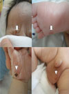
The baby size were measured length of 50 cm (50-75th percentile), a weight of 3,800 g (90th percentile), and a head circumference of 40.5 cm (75-90th percentile). The vital signs were also recorded with a blood pressure of 80/50 mmHg, temperature of 36.5℃, heart rate of 134 beats per minute, and a respiratory rate of 56 breaths per minute. Edema was noted in both lower extremities, there were multiple ulcerative plaques with crust and extensive desquamation on the face and right hand, and various erythematous maculopapular rashes on the soles and upper thighs. A number of ulcerative papules and crusted vesicles were noted, particularly on the scrotum (Fig. 1). Auscultation of the lungs revealed coarse breathing sounds with crackles. There were no signs of hepatosplenomegaly.
The laboratory test results indicated a white blood cell count (WBC) of 26,040/mm3 (5,000-19,500/mm3) with 55% polymorphonuclear leukocytes, 30% lymphocytes, 11% monocytes, 4% eosinophils, a hemoglobin level of 11.4 g/dL, and a platelet count of 488,000/mm3. Blood urea nitrogen (BUN), creatinine, alanine aminotransferase, aspartate aminotransferase and total bilirubin levels were normal.
C-reactive protein levels were elevated to 20.3 mg/L (0-5 mg/L), and a total protein level of 3.7 g/dL (4.6-7.4 g/dL) with 1.8 g/dL (3.9-5.0 g/dL) albumin indicated hypoalbuminemia. Urine analysis showed proteinuria (+++) and hematuria (+++) with 50-60% dysmorphic erythrocytes on microscopic examination. The spot urine protein/creatinine ratio was elevated to 26.86, which was in nephrotic range.
Serological tests confirmed syphilis, rapid plasma reagin (RPR) titer was 1:32.0 (<1:2) and positive for syphilitic (FTA-ABS) immunoglobulin M (IgM). The mother's RPR titer was 1:96.0 and treponemal pallidum antibody and FTA-ABS IgM was positive. Prothrombin time and partial thromboplastin time were 10.8 and 35.3 seconds respectively and were both within the normal range. The HIV serological test was negative.
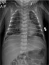
The patient's cerebrospinal fluid examination revealed values within normal limits (protein 54 mg/dL, glucose 82 mg/dL) and was negative for VDRL, bacterial and fungal culture, not including pleocytosis (white blood cell values were 22/mm3 with 90% lymphocytes). The chest radiograph revealed multiple rounded, parenchymal nodules that sprouted from the hilum and spread throughout the lung, and were notable on the base of the lung (Fig. 2). Bone radiographs of the wrists, elbows, knees and ankles showed normal findings. Abdominal ultrasonography revealed no significant hepatosplenomegaly.
The infant was diagnosed as congenital syphilis with nephrotic syndrome and pneumonia alba. He was treated with aqueous penicillin G 50,000 U/kg/dose, for 10 days at 8 hour intervals and improvements in the skin lesions, pneumonia, proteinuria, and hematuria were observed afterward. Serological and urinary inspections 2 weeks after the termination of treatment revealed normal findings. The following test results were obtained: total protein 5.1 g/dL, albumin 3.4 g/dL, RPR titier 1:2.5, FTA-ABS IgM (-) and FTA-ABS IgG (+). Clinical and laboratory findings all indicated that the patient had recovered. As soon as he diagnosed, his mother was also treated with penicillin G 2,400,000 U/dose for 14 days.
Discussion
Congenital syphilis can present with diverse clinical findings and can affect almost every organ. The symptoms are usually identified 3-4 weeks after birth. Typical clinical manifestations include rhinitis, maculopapular or exfoliative rash, hepatosplenomegaly, and jaundice. Bone involvement such as osteochondritis and periostitis, a change in cerebrospinal fluid, fever, anemia, and pneumonia alba also could be present with congenital syphilis. However, there are currently no reports about congenital syphilis with concurrent signs of nephrotic syndrome and pneumonia alba, which is described in this paper.
Cutaneous involvement in patients with congenital syphilis has been reported to occur in up to 70 percent of infants. It is characterized by a rash that covers the palms and soles that may be covered with scales and is often followed by desquamation. Syphilitic pemphigus is another typical vesicular eruption that is covered with a brown callus after the vesicle ruptures. These cutaneous lesions could result from congenital syphilis, but could also occur with Staphylococcus aureus, Pseudomonas aeruginosa, Listeria monocytogenes, Haemophilus influenzae type b, Mycobacterium tuberculosis, cytomegalovirus and herpes virus infections. Therefore a diagnosis requires histologic analysis, dark-field microscopy or an immunofluorescent microscopic test of the serum in ulcerative lesion to determine the causative organism3). The infant in this case had clinical manifestations of maculopapular eruption, vesicle, callus, and desquamation. However, histological or bacteriological tests had not been performed which limits the ability to confirm the relationship of these cutaneous lesions with congenital syphilis.
Nephrotic syndrome is a well-known complication of congenital syphilis that presents with severe proteinuria, hypoalbuminemia, hypercholesterolemia, and edema. One indication of nephrotic syndrome is when the amount of protein excreted in the urine in a 24-hour period exceeds 960 mg/m2. The spot urine protein/creatinine ratio (UPCR) in a random sample could be used as an alternative to a 24-hour sample to detect nephrotic syndrome. In this case, a 24-hour sample test was not performed and the UPCR in a random sample was 26.86 which indicated proteinuria and nephrotic syndrome was presumed. Finally, a renal biopsy was considered unnecessary in this case because congenital syphilis was confirmed with a serologic test and the patient fully recovered as a result of treatment of the primary disease.
Renal involvement is characterized by immunoglobulin, treponemal antigenic material, C3 and fibrin deposit findings in the glomerular basal membrane which indicate that the development of nephrotic syndrome is caused by an immune complex secondary to syphilis. A study of 455 infants with congenital syphilis Thailand showed that 11 cases (2.4%) had nephrotic syndrome, and two of these cases died because of necrotizing enterocolitis and sepsis (mortality rate 18%)4). Another study in Brazil identified nephrotic syndrome in 2 (5.3%) of 38 infants that were diagnosed with congenital syphilis at birth or within the first 4 weeks of life5). Appropriate penicillin and supportive therapy can allow complete clinical recovery from these conditions1, 5, 6, 7).
Respiratory distress, described as pneumonia alba, is an infrequent clinical feature of congenital syphilis. A plain chest radiographic examination revealed interstitial infiltrations with diffuse rough nodular variates to band-like opacities that radiated from the hilum8). This radiographic appearance, however, was nonspecific and a supplemental chest high resolution computed tomography (HRCT) could be performed to specify and locate the size and area of the lesion for accurate diagnosis. In this case, a similar radiographic pattern of multiple nodules that radiated from the hilum was observed on a plain chest x-ray; both the clinical symptoms and radiographic abnormalities resolved after penicillin therapy was initiated, which suggested pneumonia alba. There were no chest HRCT and supplemental inspections for other possible causes of pneumonia in this case. Pneumonia alba concurrent with congenital syphilis, is primarily reported to involve neonatal respiratory distress syndrome which can be diagnosed with nonspecific diffuse pulmonary involvement on a plain chest X-ray. However, such findings and no other discernible etiology can be considered as congenital syphilis and HRCT can be used for differential diagnosis2).
In Korea, congenital syphilis cases have been reported occasionally and combined pneumonia is not rare. A study of 32 infants with congenital syphilis showed that 38% had pneumonia9). In another clinical observation of 84 cases of congenital syphilis, pneumonia was combined in 10 cases10). There are a few reports on renal involvement in congenital syphilis in Korea. There was a case of congenital syphilis nephritic syndrome in a 2 month-old infant who recovered after penicillin therapy11). In a study of 46 cases of syphilis, during the treatment one case had nephrotic syndrome and another had acute pyelonephritis12). Thus, this case report is meaningful because there has never been reported congenital syphilis with pneumonia and nephrotic syndrome.
Syphilis has been a recognized and diagnosed infection for centuries and remains a worldwide public health problem. The rates of syphilis diminished with the use of Penicillin in the early 1940s, however, in recent years it has re-emerged, predominantly in developing countries that have not eradicated congenital syphilis. According to the USA Centers for Disease Control and Prevention, the incidence of congenital syphilis has increased from 8.2 cases in 2005 to 10.1 cases in 2008 per 100,000 surviving babies. In addition, Surveillance for Sexually Transmitted Disease has reported an increased incidence of congenital syphilis from 3 cases in 2002 to 8.7 cases in 2011 per 100,000 births in the state of Ohio, USA. Clinical findings may be subtle in neonates, which can delay recognition and diagnosis, therefore, complete prenatal screening is critical for congenital syphilis prevention13, 14). Considering the diagnostic period of this case, antenatal inspection for syphilis in developing countries is strongly encouraged. Currently, pregnant women are routinely tested for syphilis at the beginning of pregnancy, but those who are at high risk for syphilis are advised to take an additional test during the third trimester. Due to the re-emergence of syphilis and congenital syphilis, the World Health Organization recommends antenatal screening during the first and third trimester for every pregnant woman. The infant in this case was delivered by a mother that had not received appropriate prenatal care. The patient presented with cutaneous lesions with typical features such as nephrotic syndrome and pneumonia, and in similar cases, a serologic test should be performed immediately to determine a differential diagnosis. Finally, particular attention should be given to treating not only the infant but mothers that are also infected.
 XML Download