Abstract
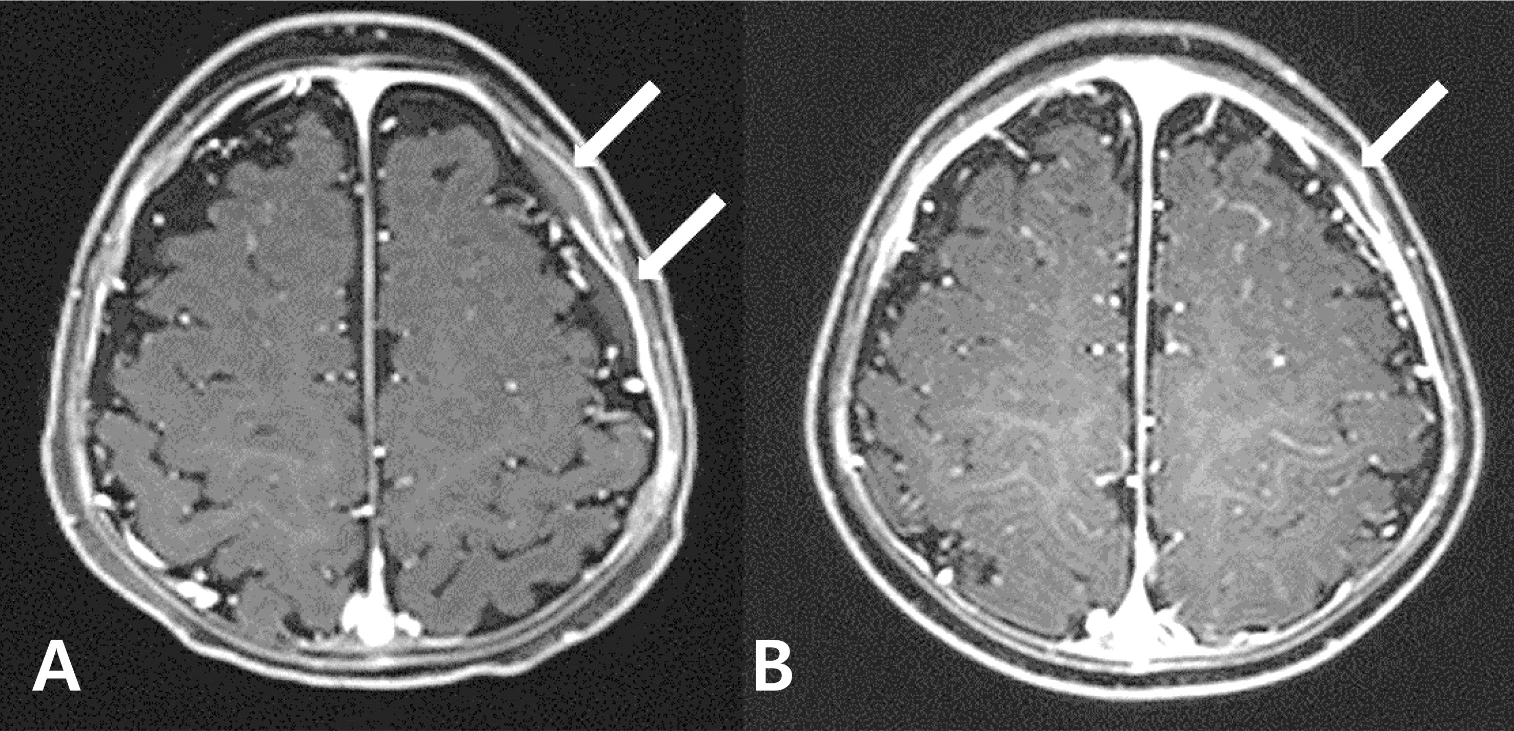
Cases of incomplete Kawasaki disease (KD), wherein the patient does not fulfill the full diagnostic criteria for KD, are often detected in infants younger than 6 months of age. The clinical manifestations in infants with incomplete KD may resemble other infectious diseases, including meningitis. For this reason, clinicians may have difficulty differentiating incomplete KD from other infectious diseases in this population. Various neurological features are associated with KD, including aseptic meningitis, subdural effusion, facial nerve palsy, cerebral infarction, encephalopathy, and reversible corpus callosum splenial lesions on magnetic resonance imaging. We report a case of a 5-month-old girl with incomplete KD, associated with cerebrospinal fluid pleocytosis and an epidural fluid collection. Echocardiography indicated dilatation of the main coronary arteries. The girl made a complete recovery, with resolution of both the epidural fluid collection and coronary artery aneurysms. In this case, the child is well, and showed normal developmental milestones at the 7-month follow-up.
REFERENCES
2. Rowley AH, Gonzalez-Crussi F, Gidding SS, Duffy CE, Shulman ST. Incomplete Kawasaki disease with coronary artery involvement. I Pediatr. 1987; 110:409–13.

3. Yeom IS, Woo HO, Park IS, Park ES, 860 IH, Youn HS. Kawasaki disease in infants. Korean I Pediatr. 2013; 56:377–232.

4. Cabral M, Correia P, Brito M], Conde M, Carreiro H. Kawasaki disease in a young infant: diagnostic challenges. Acta Reumatol Port. 2011; 36:304–8.
5. Fujiwara S, Yamano T, Hattori M, Fujiseki Y, Shimada M. Asymptomatic cerebral infarction in Kawasaki disease. Pediatr Neurol. 1992; 8:235–6.

6. Tabarki B, Mahdhaoui A, Selmi H, Yacoub M, Essoussi AS. Kawasaki disease with predominant central nervous system involvement. Pediatr Neurol. 2001; 25:239–41.

7. Takanashi I, Shirai K, Sugawara Y, Okamoto Y, Obonai T, Terada H. Kawasaki disease complicated by mild encephalopathy with a reversible splenial lesion (MERS). J Neurol Sci. 2012; 315:167–9.

8. Bailie NM, Hensey 0}, Ryan S, Allcut D, King MD. Bilateral subdural collections-an unusual feature ofpossible Kawasaki disease. Eur I Paediatr Neurol. 2001; 5:79–81.
9. Limbach HG, Lindinger A. Kawasaki syndrome in infants in the first 6 months of life. Klin Padiatr. 1991; 203:133–6.
10. Chang FY, Hwang B, Chen S], Lee PC, Meng CC, Lu IH. Characteristics of Kawasaki disease in infants younger than six months of age. Pediatr Infect Dis I. 2006; 25:241–4.

11. Yeom IS, Park IS, Seo IH, Park ES, Lim IY, Park CH, et al. Initial characteristics of Kawasaki disease With cerebrospinal fluid pleocytosis in febrile infants. Pediatr Neurol. 2012; 47:259–62.
12. Dengler LD, Capparelli EV, Bastian IF, Bradley D], Glode MP, Santa S, et al. Cerebrospinal fluid profile in patients with acute Kawasaki disease. Pediatr Infect Dis I. 1998; 17:478–81.
13. Terasawa K, Ichinose E, Matsuishi T, Kato H. Neurological complications in Kawasaki disease. Brain Dev. 1983. 5z371–4.

14. Takagi K, Umezawa T, Saji T, Morooka K, Matsuo N. Meningoencephalitis in Kawasaki disease. No To Hattatsu. 1990; 22:429–35.


 PDF
PDF ePub
ePub Citation
 Print
Print


 XML Download