Abstract
Macrophage activation syndrome (MAS) is a rare complication in systemic lupus erythematosus (SLE) that can be triggered by infections. Due to the fact that MAS may mimic clinical features of underlying rheumatic disease, or be confused with an infectious complication, its detection can prove challenging. This is particularly true when there is an unknown/undiagnosed disease; and could turn into an even greater challenge if MAS and SLE are combined with a viral infection. A—i4-year—old female came to the hospital with an ongoing fever for 2 weeks and a painful facial skin rash. Hepatomegaly, pancytopenia, increased aspartate aminotransferase, elevated serum ferritin and lactate dehydrogenase were reported. No hemophagocytic infiltration of bone marrow was reported. The patient was suspected for hemophagocytic lymphohistiocytosis. Her skin rashes were eczema herpeticum, which is usually associated with immune compromised conditions. With the history of oral ulcers and malar rash, positive ANA and low C3, C4 and the evidence of hemolytic anemia, she was diagnosed as SLE. According to the diagnostic guideline for MAS in SLE, she was diagnosed MAS as well, activated by acute HSV infection. After administering steroids and antiviral agent, the fever and skin rash disappeared, and the abnormal laboratory findings normalized. Therefore, we are reporting a rare case of MAS triggered by acute HSV infection as the first manifestation of SLE.
REFERENCES
1. Stichweh D, Arce E, Pascual V. Update on pediatric systemic lupus erythematosus. Curr Opin Rheumatol. 2004; 16:577–87.

2. Avcin T, Tse SM, Schneider R, Ngan B, Silverman ED. Macro—phage activation syndrome as the presenting manifestation of rheumatic diseases in childhood. I Pediatr. 2006; 148:683–6.
3. Shimizu M, Yokoyama T, Tokuhisa Y, Ishikawa S, Sakakibara Y, Ueno K, et al. Distinct cytokjne profile in juvenile systemic lupus erythematosus-associated macrophage activation syndrome. Clin Immunol. 2013; 146:73–6.
4. Parodi A, Davi S, Pringe AB, Pistorio A, Ruperto N, Magni-Manzoni S, et al. Macrophage activation syndrome in juve—nile systemic lupus erythematosus: a multinational mul—ticenter study of thirty—eight patients. Arthritis Rheum. 2009; 60:3388–99.

5. Ramos—Casals M, Cuadrado M}, Alba P, Sanna G, Brito—Zerén P, Bertolaccini L, et al. Acute viral infections in patients with systemic lupus erythematosus: description of 23 cases and review of the literature. Medicine. 2008; 87:311–8.
6. Ueda Y, Yamashita H, Takahashi Y, Kaneko H, Kano T, Mi—mori A. Refractory hemophagocytic syndrome in systemic lupus erythematosus successfully treated with intermittent intravenous cyclophosphamide: three case reports and 1i—terature review. Clin Rheumatol. 2014; 33:281–6.
7. Petri M, Orbai AM, Alarcon GS, Gordon C, Merrill IT, Fortin PR, et al. Derivation and validation of the Systemic Lupus International Collaborating Clinics classification criteria for systemic lupus erythematosus. Arthritis Rheum. 2012; 64:2677–86.
8. Fukaya S, Yasuda S, Hashimoto T, Oku K, Kataoka H, Horita T, et al. Clinical features of haemophagocytic syndrome in patients With systemic autoimmune diseases: analysis of 30 cases. Rheumatology (Oxford). 2008; 47:1686–91.

9. Egfies Dubuc CA, Uriarte Ecenarro M, Meneses Villalba C, Aldasoro Céceres V, Hernando Rubio I, Belzunegui Otano I. Hemophagocytic syndrome as the initial manifestation of systemic lupus erythematosus. Reumatol Clin. 2014; 10:321–4.
10. Jiménez AT, Vallejo ES, Cruz MZ, Cruz AC, Jara BS. Mac—rophage activation syndrome as the initial manifestation of severe juvenile onset systemic lupus erythematosus. Favorable response to cyclophosphamide. Reumatologia Clinica (English Edition). 2014; 10:331–5.
11. Vilaiyuk S, Sirachainan N, Wanitkun S, Pirojsakul K, Vaew—panich I. Recurrent macrophage activation syndrome as the primary manifestation in systemic lupus erythematosus and the benefit of serial ferritin measurements: a case-based re—view. Clin Rheumatol. 2013; 32:899–904.

12. Stephan I, Koné—Paut I, Galambrun C, Mouy R, Bader—Meu—nier B, Prieur AM. Reactive haemophagocytic syndrome in children with inflammatory disorders. A retrospective study of 24 patients. Rheumatology. 2001; 40:1285–92.
13. Yeap ST, Sheen IM, Kuo HC, Hwang KP, Yang KD, Yu HR. Macrophage activation syndrome as initial presentation of systemic lupus erythematosus. Pediatr Neonatol. 2008; 49:39–42.

14. Lambotte O, Khellaf M, Harmouche H, Bader—Meunier B, Manceron V, Goujard C, et al. Characteristics and long—term outcome of 15 episodes of systemic lupus erythematosus-associated hemophagocytic syndrome. Medicine (Baltimore). 2006; 85:169–82.

15. Sawhney 8, Woo P, Murray K]. Macrophage activation synd—rome: a potentially fatal complication of rheumatic disorders. Arch Dis Child. 2001; 85:421–6.
16. Isome M, Suzuki 1, Takahashi A, Murai H, Nozawa R, Suzuki S, et al. Epstein-Barr Virus—associated hemophagocytic syndrome in a patient with lupus nephritis. Pediatr Nephrol. 2005; 20:226–8.

17. Behrens EM, Beukelman T, Paessler M, Cron RQ. Occult macrophage activation syndrome in patients with systemic juvenile idiopathic arthritis. I Rheumatol. 2007; 34:1133–8.
18. Benarroch LK, Sterba G, Bosque M. Macrophage activation syndrome (MAS) as a debut of systemic lupus erythematous (SLE) in a child. I Allergy Clin Immunol. 2006; 117:8210.
19. Hur M, Kim YC, Lee KM, Kim KN. Macrophage activation syndrome in a child with systemic juvenile rheumatoid arthritis. I Korean Med Sci. 2005; 20:695–8.

20. Shim YS, Kim HS, Kim KN. A case of macrophage activation syndrome successfully treated with combination therapy including etanercept. I Rheum Dis. 2012; 19:225–9.
21. Keum SW, Kim M], Bae EY, Ham SB, Chung NK, Ieong DC, et al. A case of macrophage activation syndrome developed in female adolescent with systemic lupus erythematosus. I Rheum Dis. 2014; 21:96–100.

22. Hwang JY, No SM, Lee I, Iang PS, Kim YH, Kim IT, et al. A case of hemophagocytic lymphohistiocytosis in a child with systemic lupus erythematosus. Korean I Pediatr. 2003; 46:1029–31.
23. Kim ES, Kim YG, Kim WS, Jung YS, Han JH, Bae CB, et al. Three cases of secondary hemophagocytic lymphohistiocytosis associated with systemic erythematosus lupus. I Rheum Dis. 2015; 22:180–5.
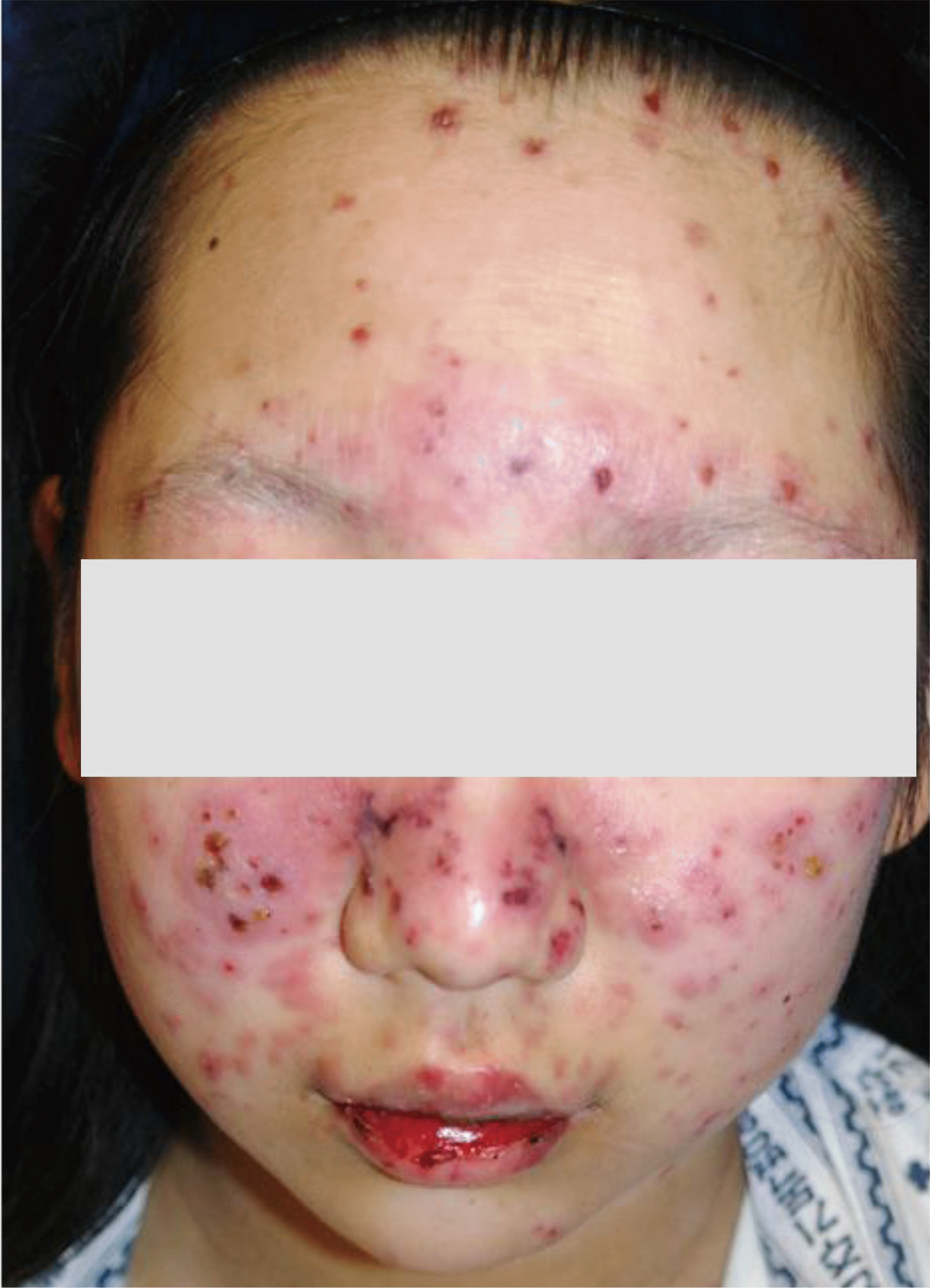
Fig. 1.
Multiple diffuse erythematous papules, pustules, and patches with edema were on the face and scalp.


 PDF
PDF ePub
ePub Citation
 Print
Print


 XML Download