Abstract
Purpose
Osteoarticular infections in children and adolescents are important because it can cause functional compromise if appropriate treatment is delayed. Therefore, this study was designed to describe the clinical presentations and causative organisms of osteoarticular infections in children and adolescents in order to propose early diagnosis method and an appropriate empiric antimicrobial therapy.
Methods
Forty-two medical records were reviewed retrospectively, which were confirmed as osteomyelitis (OM) 0r septic arthritis (SA) at Department of Pediatrics 0r Orthopedic Surgery in patients under 18 years old of Ewha Womans University Mokdong Hospital from March 2008 to March 2015.
Results
We identified 21 cases of OM, 13 cases of SA and 8 cases of OM with SA. There were 31 males and 11 females and mean age was 7.1 years old. The most common symptoms were pain and tenderness of involved site. Major involved bones were femur (10 cases, 34.5%), tibia (7 cases, 24.1%) and major involved joints were hip (9 cases, 42.9%), and knee (5 cases, 23.8%). Increased serum C-reactive protein and erythrocyte sedimentation rate were observed in 37 cases (88.1%) respectively. Magnetic resonance imaging was performed in 40 cases among 42 cases and was used to demonstrate osteoarticular infections and other adjacent infections. Nine cases (23.7%) among 38 cases and 20 cases (50.0%) among 40 cases were positive in blood culture and infected site culture respectively. The most common causative organism was StaphyLococcus aureus, which was represented in 22 cases (75.9%), of which nine cases (40.9%) were resistant to methicillin.
Conclusions
S. aureus was the most common causative organism of osteoarticular infections in children and adolescents and the proportion of MRSA was high in this study. Therefore, we recommend vancomycin as the first empiric antimicrobial therapy and suggest that further study is necessary to elucidate an appropriate guideline for treatment which takes into account MRSA proportion.
Go to : 
REFERENCES
1. Bonhoeffer I, Haeberle B, Schaad UB, Heininger U. Diagnosis of acute haematogenous osteomyelitis and septic arthritis: 20 years experience at the University Children's Hospital Basel. Swiss Med Wkly. 2001; 131:575–81.
3. Paakkonen M, Peltola H. Bone and joint infections. Pediatr Clin North Am. 2013; 60:425–36.
4. Chen WL, Chang WN, Chen YS, Hsieh KS, Chen CK, Peng N], et al. Acute community-acquired osteoarticular infections in children: high incidence of concomitant bone and joint involvement. I Microbiol Immunol Infect. 2010; 43:332–8.

5. Dodwell ER. Osteomyelitis and septic arthritis in children: current concepts. Curr Opin Pediatr. 2013; 25:58–63.
6. Russell CD, Ramaesh R, Kalima P, Murray A, Gaston MS. Microbiological characteristics of acute osteoarticular infections in children. I Med Microbiol. 2015; 64:446–53.

7. Williams D]. Deis IN, Tardy I, Creech CB. Culture-negative osteoarticular infections in the era of community-associated methicillin-resistant Staphylococcus aureus. Pediatr Infect Dis 12011305235.
8. Goergens ED, McEvoy A, Watson M, Barrett IR. Acute osteomyelitis and septic arthritis in children. J Paediatr Child Health. 2005; 41:59–62.

9. Montgomery NI, Rosenfeld S. Pediatric osteoarticular infection update. I Pediatr Orthop. 2015; 35:74–81.

10. Grammatico-Guillon L, Maakaroun Vermesse Z, Baron S, Gettner S, Rusch E, Bernard L. Paediatric bone and joint infections are more common in boys and toddlers: a national epidemiology study. Acta Paediatr. 2013; 102:e120–5.

11. Choi IH, Choe Yj, Hong KB, Lee I, Yoo W], Kim HS, et al. The etiology and clinical features of acute osteoarthritis in children; 2003—2009. Korean I Pediatr Infect Dis. 2011; 18:31–9.

12. Gillespie W]. Epidemiology in bone and joint infection. Infect Dis Clin North Am. 1990. 4z361–76.
13. Rasmont Q, Yombi IC, Van der Linden D, Docquier PL. Osteoarticular infections in Belgian children: a survey of clinical, biological, radiological and microbiological data. Acta Orthop Belg. 2008; 74:374–555.
14. Paakkonen M, Kallio Mj, Kallio PE, Peltola H. Sensitivity of erythrocyte sedimentation rate and C-reactive protein in childhood bone and joint infections. Clin Orthop Relat Res. 2010; 468:861–6.
15. Unkila-Kallio L, Kallio Mj, Eskola I, Peltola H. Serum C-reactive protein, erythrocyte sedimentation rate, and white blood cell count in acute hematogenous osteomyelitis of children. Pediatrics. 1994; 93:59–62.

16. Riise OR, Kirkhus E, Handeland KS, Flato B, Reiseter T, Cvancarova M, et al. Childhood osteomyelitis-incidence and differentiation from other acute onset musculoskeletal features in a population-based study. BMC Pediatr. 2008. 8245.

17. Butbul-Aviel Y, Koren A, Halevy R, Sakran W. Procalcitonin as a diagnostic aid in osteomyelitis and septic arthritis. Pediatr Emerg Care. 2005; 21:828–32.

18. Park SS, Lee SH, Sim GB. MRI in suspected acute septic arthritis of the hip joint in children. Hip Pelvis. 2012; 24:295–301.

19. Levy PY, Fournier PE, Fenollar F, Raoult D. Systematic PCR detection in culture-negative osteoarticular infections. Am J Med. 2013; 126:1143.e25–33.

20. Ceroni D, Cherkaoui A, Ferey S, Kaelin A, Schrenzel I. KingeHa kingae osteoarticular infections in young children: clinical features and contribution of a new specific realtime PCR assay to the diagnosis. J Pediatr Orthop. 2010; 30:301–4.
21. Dubnov-Raz G, Ephros M, Garty BZ, Schlesinger Y, Maayan-Metzger A, Hasson I, et al. Invasive pediatric KingeHa kingae infections: a nationwide collaborative study. Pediatr Infect Dis J. 2010; 29:639–43.
22. Dubnov-Raz G, Scheuerman O, Chodick G, Finkelstein Y, Samra Z, Garty BZ. Invasive KingeHa kingae infections in children: clinical and laboratory characteristics. Pediatrics. 2008; 122:1305–9.
23. Park IH, Lee T]. Increasing rates of community associated methicillin-resistant Staphylococcus aureus in children with muscular-skeletal infections in Korea: a single center experience from 2000 to 2012. Korean I Pediatr Infect Dis. 2013; 20:63–70.
24. Kwak YH, Park SE, Hong IY, Jung HS, Park IY, Choi IH, et al. Etiologic agents and clinical features of acute pyogenic osteoarthritis in children. I Korean Pediatr. 2000; 43:506–13.
25. Koo M, Kim D. The clinical aspects of septic arthritis in children. I Korean Pediatr. 1997; 40:1737–44.
Go to : 
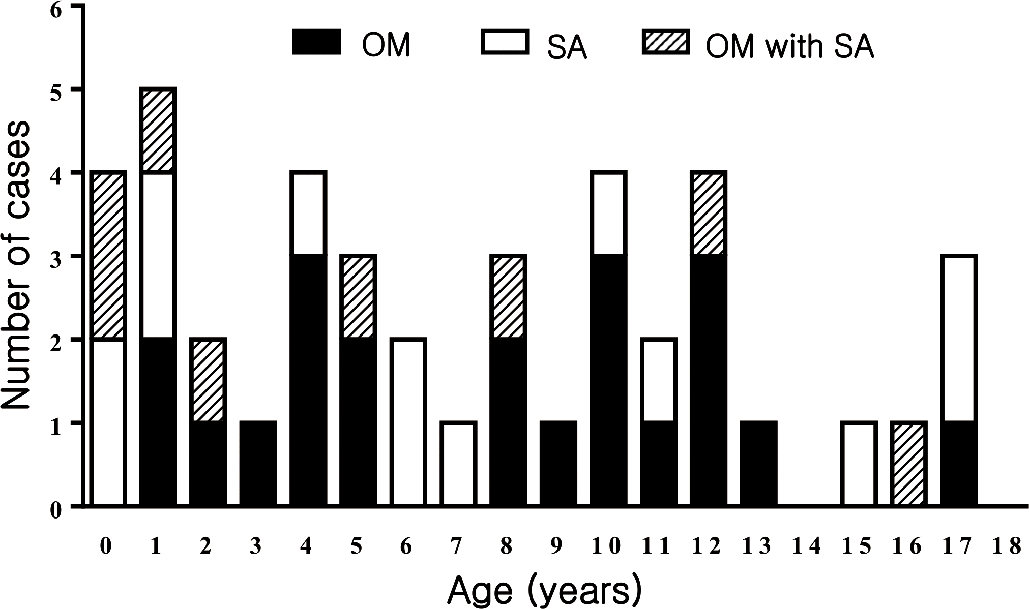
 | Fig. 1.Age distribution of 42 children and adolescents with osteoarticular infections. Number of cases of osteomyelitis (0M), septic arthritis (SA) and osteomyelitis with septic arthritis were arranged by age of diagnosis. |
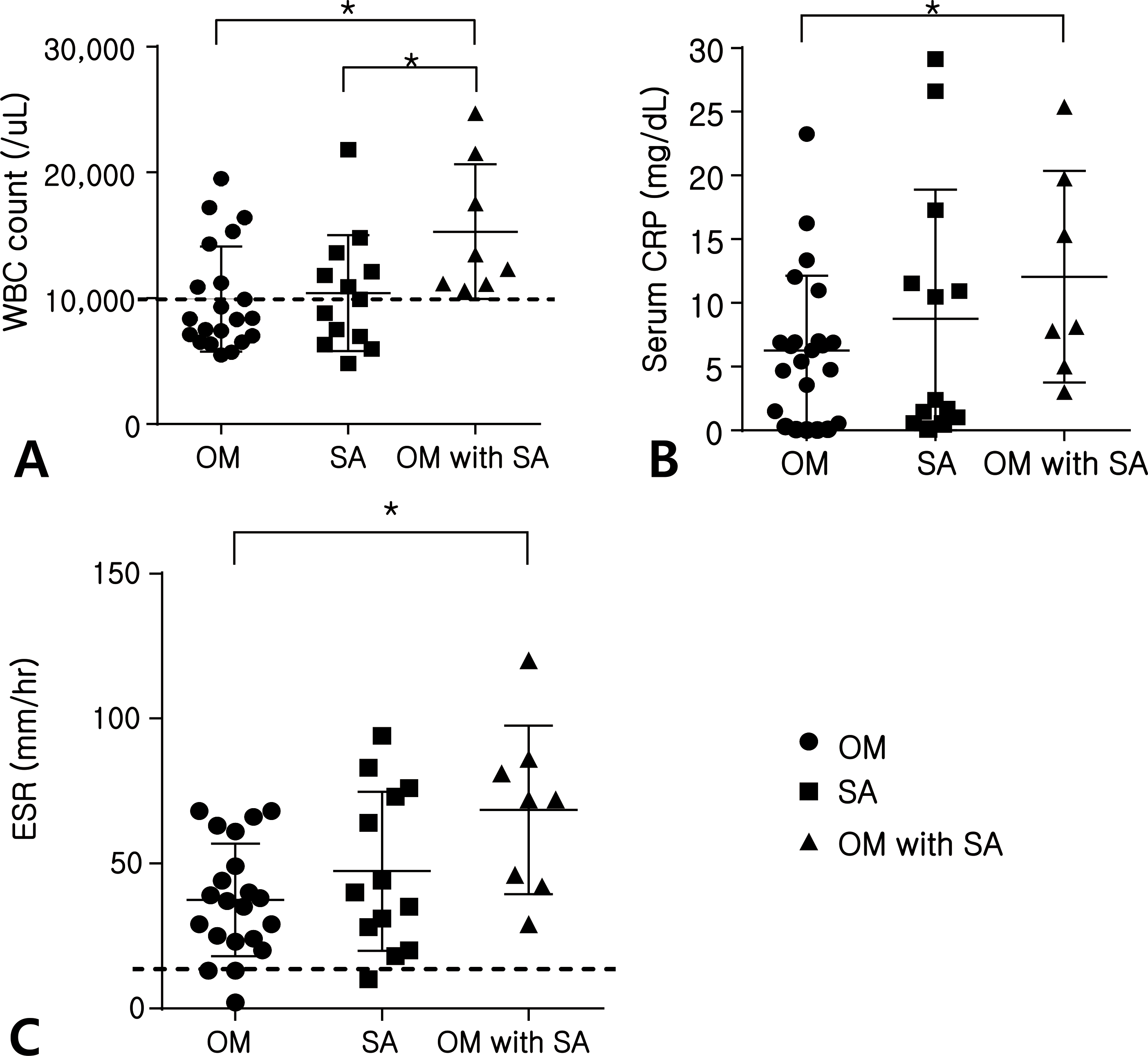
 | Fig. 2.Inflammatory biomarkers of blood in children and adolescents with osteoarticular infections. White blood cell (WBC) count (A), concentration of C-reactive protein (CRP) (B) and erythrocyte sedimentation rate (ESR) (C) were compared in children and adolescents with osteomyelitis (0M), septic arthritis (SA) and osteomyelitis with septic arthritis (∗P<0.05; means upper normal of WBC count or ESR; 0M, osteomyelitis; SA, septic arthritis). |
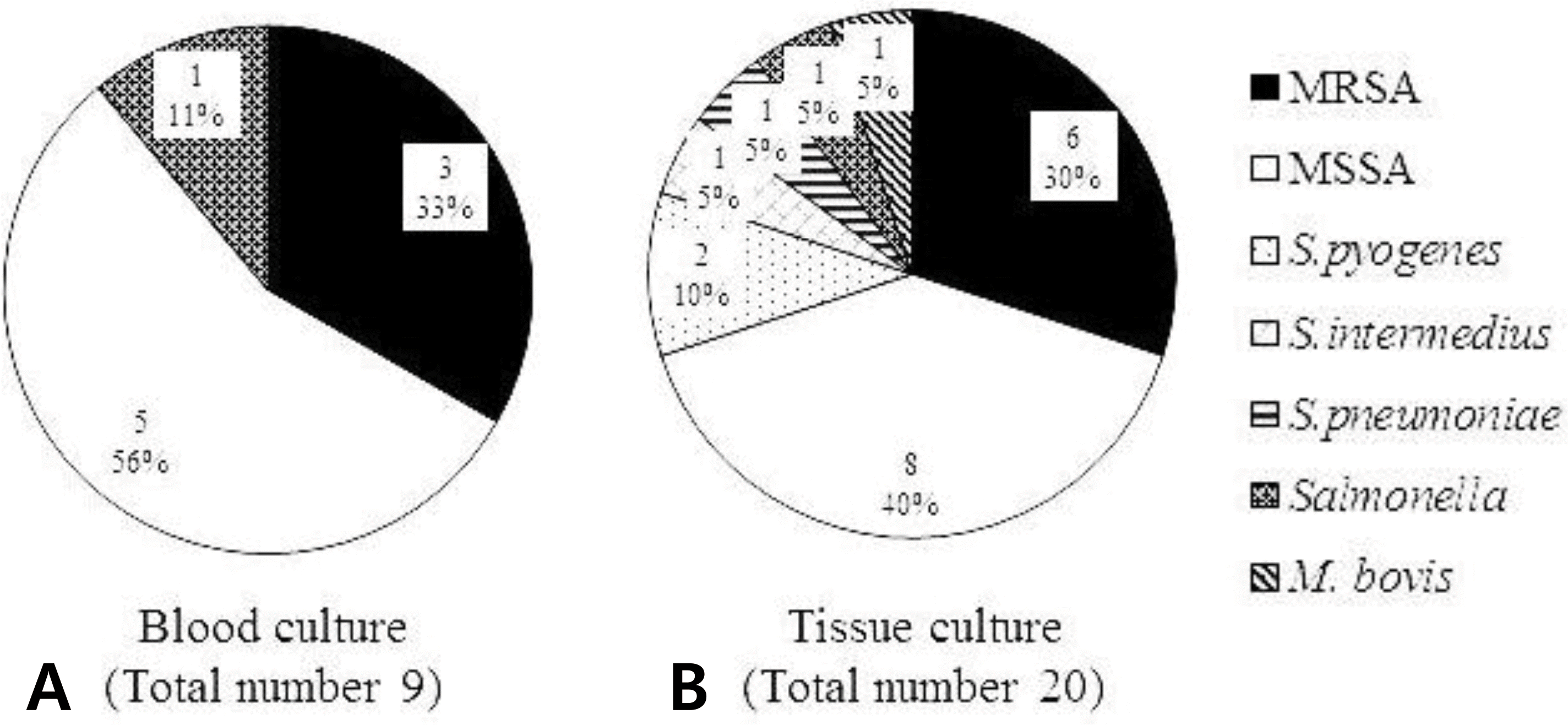
 | Fig. 3.Isolated causative organisms from blood (A) and infected tissue culture (B) from children and adolescents with osteoarticular infections. Causative organisms were isolated from 9 cases among 38 cases of blood culture (A) and from 20 cases among 40 cases of tissue or synovial fluid culture (B). The most common causative organism was S. aureus in both cultures (MRSA, methicillin-resistant Staphylococcus aureus; MSSA, methicillin- sensitive Staphylococcus aureus; 0M, osteomyelitis; SA, septic arthritis). |
Table 1.
Clinical Characteristics, Laboratory and Radiologic Findings in Children and Adolescents with Ostearticular ln-fections
Table 2.
Involved Sites in Children and Adolescents with Osteoarticular Infections


 PDF
PDF ePub
ePub Citation
 Print
Print


 XML Download