Abstract
Infantile osteopetrosis is a rare congenital disorder caused by abnormal bone resorption. Patients with osteopetrosis can have severe anemia, thrombocytopenia, hepatosplenomegaly, rickets, visual impairment, and deafness. Cytomegalovirus also can cause a congenital infection with anemia, thrombocytopenia, hepatosplenomegaly, and calcifications in the brain. We report a 38-day-old infant with severe hepatosplenomegaly, thrombocytopenia, hypocalcemia, and growth failure. Real time polymerase chain reaction detected cytomegalovirus in the plasma. Skeletal radiography revealed generalized bone sclerosis. He was diagnosed with osteopetrosis along with cytomegalovirus infection. Only the test for mutation of the CLCN7 gene, representing the most common and heterogeneous form of osteopetrosis, was available, and the result was negative. With supportive care and antiviral treatment, severe thrombocytopenia due to the cytomegalovirus infection almost normalized despite the possible immunosuppression caused by osteopetrosis. We present the first report of an infant who suffered from osteopetrosis and CMV infection which was successfully treated by long term antiviral agent therapy.
REFERENCES
1. Mazzolari E, Forino C, Razza A, Porta F, Villa A, Notaran-gelo LD. A single-center experience in 20 patients with infantile malignant osteopetrosis. Am J Hematol. 2009; 84:473–9.

3. Siddaiahgari SR, Makadia D, Shah N, Devi RR, Lingappa L. Identification of novel mutation in autosomal recessive infantile malignant osteopetrosis. Indian I Pediatr. 2014; 81:969–70.

4. Sobacchi C, Schulz A, Coxon FP, Villa A, Helfrich MH. Osteopetrosis: genetics, treatment and new insights into osteoclast function. Nat Rev Endocrinol. 2013. 9z522–36.

5. Nyholm IL, Schleiss MR. Prevention of maternal cytomegalovirus infection: current status and future prospects. Int I Womens Health. 2010; 2:23–35.
6. Ihde LL, Forrester DM, Gottsegen CI, Masih S, Patel DB, Vachon LA, et al. Sclerosing bone dysplasias: review and differentiation from other causes of osteosclerosis. Radiographics. 2011; 31:1865–82.

7. Albers-Schénberg HE. Réntgenbilder einer seltenen Kno-chenerkrankung. Munch Med Wochenschr. 1904; 51:365–8.
8. Superti-Furga A, Unger S. Nosology and classification of genetic skeletal disorders: 2006 revision. Am I Med Genet A. 2007. 143A21–18.

9. Ornoy A, Diav-Citrin 0. Fetal effects of primary and secondary cytomegalovirus infection in pregnancy. Reprod Toxicol. 2006; 21:399–409.
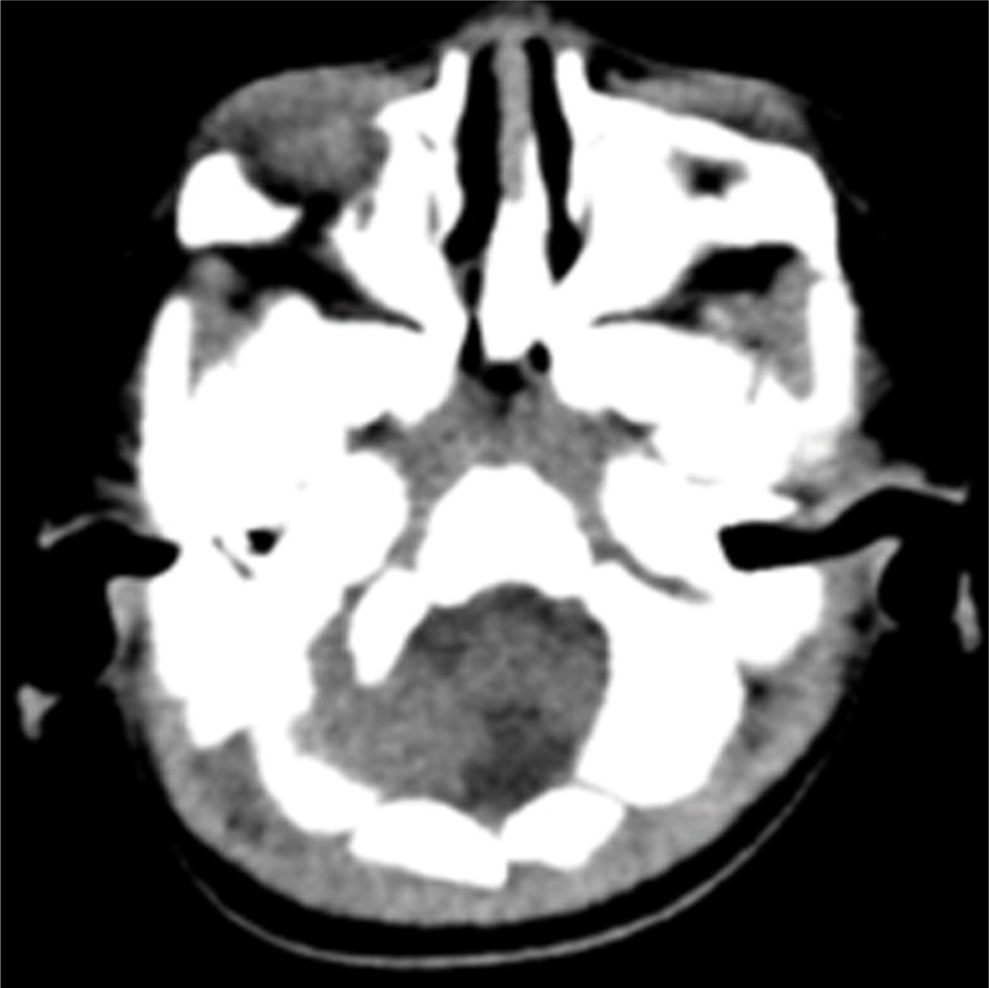
Fig. 1.
Osteopetrosis on brain computed tomography. No evidence of intracranial calcification is observed. Sclerotic change of the skull could represent osteopetrosis.

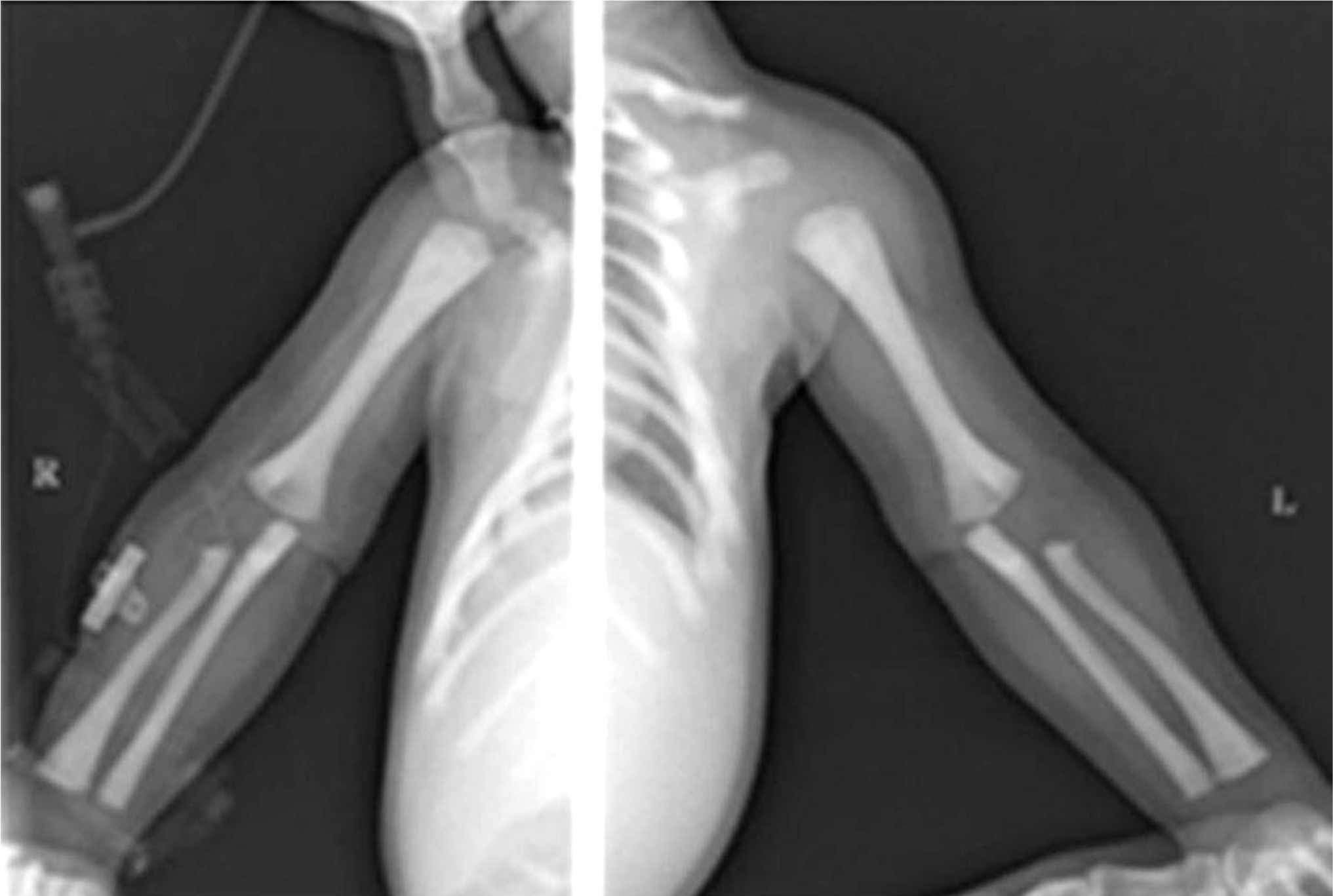
Fig. 2.
Osteopetrosis on extremity radiography. Metaphyseal flaring and increased bone density of long bones is consistent with osteopetrosis.


 PDF
PDF ePub
ePub Citation
 Print
Print


 XML Download