

 PDF
PDF ePub
ePub Citation
 Print
Print


Abstract
Erythema nodosum (EN) is a painful skin disease characterized by erythematous tender nodules located predominantly over the extensor aspects of the legs. Various etiological factors, including infection, drug administration, and systemic illness have been implicated as causes of EN. Mycoplasma pneumoniae is one of rare infectious agents to cause EN in children. We report a case of a 7-year-old boy with context of respiratory illness and skin lesions with arthralgia. From stepwise approaches, IgM antibody against M. pneumoniae was positive with titers of 12.18, consistent with respiratory infection of M. pneumoniae and histopathology showed findings of septal and lobular inflammation without vasculitis consistent with EN. In addition, we reviewed the pathogenesis of this disease based on our case and the previous reports.
REFERENCES
1. Polcari IC, Stein SL. Panniculitis in childhood. Dermatol Ther. 2010; 23:356–67.

2. Cox NH, Jorizzo IL, Bourke IF, Savage COS. Vasculitis, neutrophilic dermatoses and related disorders. Burns T, Breathnach S, Cox N, Grifliths C, editors. editors.Rook's textbook of dermatology. 8th ed.Oxford: Wiley-Blackwell;2010. p. 1282–7.
3. Kakourou T, Drosatou P, Psychou F, Aroni K, Nicolaidou P. Erythema nodosum in children: a prospective study. I Am Acad Dermat01. 2001; 44:17–21.
4. Rodriguez Blanco MA, Martin Morales IM, Casas Fernandez L. Erythematoviolaceous nodules and pneumonia. An Esp Pediatr. 2002; 57:279–80.
5. Shimizu M, Hamaguchi Y, Matsushita T, Sakakibara Y, Yachie A. Sequentially appearing erythema nodosum, erythema multiforme and Henoch-Schénlein purpura in a patient With Mycoplasma pneumoniae infection: a case report. I Med Case Rep. 2012; 6:398.
6. Aydin-Teke T, Tanir G, Bayhan GI, Metin 0, Oz N. Erythema nodosum in children: evaluation of 39 patients. Turk] Pediatr. 2014; 56:144–9.
7. Greco F, Catania R, Pira AL, Saporito M, Scalora L, Aguglia MG, et al. Erythema nodosum and Mycoplasma pneumoniae infections in childhood: Further observations in two patients and a literature review. I Clin Med Res. 2015. 72274–7.
8. Blake T, Manahan M, Robins K. Erythema nodosum-a review of an uncommon panniculitis. Dermatology Online Journal. 2014; 20:22376.
9. Sanchez-Vargas FM, Gomez-Duarte OG. Mycoplasma pneumoniae-an emerging extra-pulmonary pathogen. Clin Microbiol Infect. 2008; 14:105- 17.
10. Schalock PC, Dinulos JG. Mycoplasma pneumoniae-induced cutaneous disease. Int I Dermatol. 2009; 48:673–80.
11. Narita M. Pathogenesis of extrapulmonary manifestations of Mycoplasma pneumoniae infection with special reference to pneumonia. J Infect Chemother. 2010; 16:162–9.
12. Kano Y, Mitsuyama Y, Hirahara K, Shiohara T. Mycoplasma pneumoniae -infectioninduced erythema nodosum, ana-phylactoid purpura, and acute urticaria in 3 people in a single family. I Am Acad Dermatol. 2007; 57:33–5.
13. Lee KY. A common immunopathogenesis mechanism for infectious diseases: the protein-homeostasis-system hypo-thesis. Infect Chemother. 2015; 47:12–26.
14. Schwartz RA, Nervi S]. Erythema nodosum: a sign of systemic disease. Am Fam Physician. 2007; 75:695–700.
Fig. 1.
Scattered erythematous subcutaneous nodules located on pretibial surface of lower extremities.
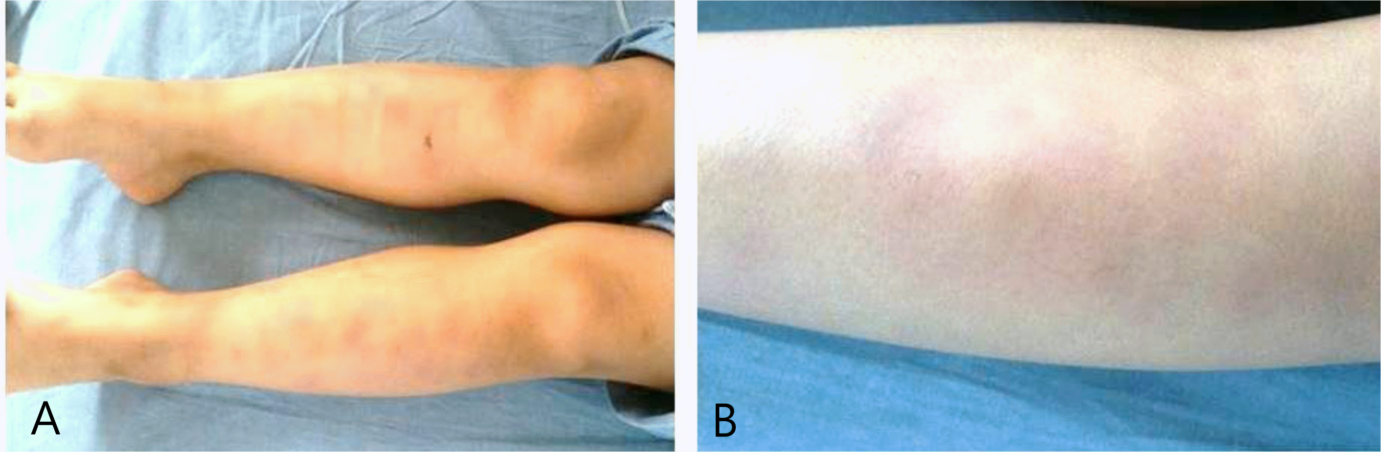
Fig. 2.
There is septal (thick arrow) and lobular (thin arrow) inflammation composed of lymphocytes mixed with histiocytes. (A, H&E stain, X 40). There is a non-caseating small granuloma (circle) composed of ephithelioid histiocytes and surrounded by lymphocytes at septa of adipose tissue. No vasculitis is observed. (B, H&E stain, X 200).
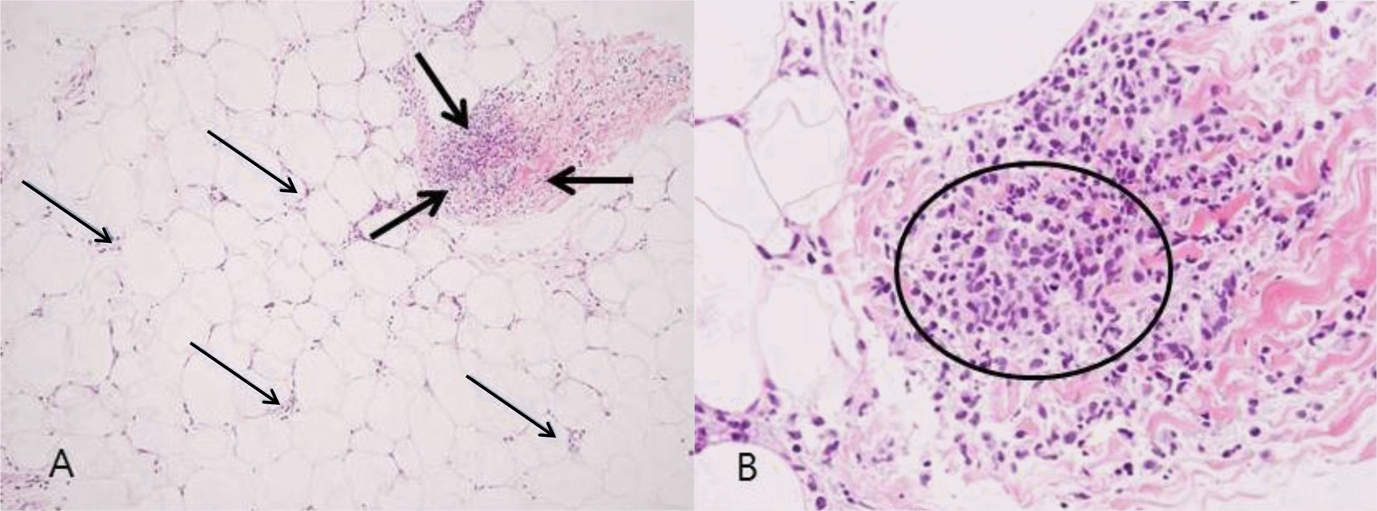
Table 1.
Summary of Mycoplasma pneumoniae Induced EN Cases in Enalish Literature;
 XML Download