

 PDF
PDF ePub
ePub Citation
 Print
Print


Introduction
Kawasaki disease (KD) is an acute self-limited systemic inflammation of unknown etiology. The major complication of KD, i.e., coronary artery lesions (CALs), became the most common cause of acquired heart disease in developed countries12). CALs occurred in 15–25% of children with KD in the era of no intravenous immunoglobulin (IVIG) treatment, but occurred in 5% of children treated with standard IVIG treatment34).
Clinical diagnostic criteria of KD include oral mucosa changes, bilateral conjunctivitis, cervical lymphadenopathy (LAP), skin rashes, and extremity changes. It was previously proposed that these signs of systemic inflammations may be caused by the inflammatory mediators after an infection of unknown KD pathogen(s). The substances that can induce inflammation are preformed in an unknown focus, and at the time of the disease onset, they are spread and reach to various organ cells including coronary artery endothelial cells as main target cells via systemic circulation. These substances and corresponding immune cells may be responsible for the various systemic inflammations shown in KD5).
It is unknown whether the degree of clinical signs may be associated with the intensity of systemic inflammation, including the risk of CALs. Although cervical LAP is the least appearing sign, it was reported that patients with a first presentation of cervical LAP had a severe clinical course with high risk of CALs6).
Polymorphorous skin rashes appear during the acute phase of the disease. Although case-report studies reported that KD patients with atypical skin rash might be associated with CALs and IVIG treatment78), the correlation between skin rash characteristics and CALs is still unclear.
Arthritis is relatively common in KD in the pre-IVIG era9), but severe arthritis patients are rare in recent IVIG era like the incidence of CALs1011). Thus, there are few studies regarding relationship between arthritis and risk of CALs.
Many clinical and laboratory parameters have been identified as predictors of CALs12) such as prolonged fever, IVIG non-responsiveness, and higher C-reactive protein12). However, predictive markers cannot be universally applied because of the difference of individual immune response to the substances that induce organ cell-specific inflammation 5).
We hypothesized that severity of clinical signs such as skin lesions or arthritis might reflect the severity of systemic inflammation and subsequently the risk of CALs. We conducted this study for characteristics of KD patients with severe skin rashes or arthritis.
Materials and Methods
The subject of this study were 220 patients diagnosed with KD and treated with IVIG at Gangdong Kyung- Hee University Hospital, Korea between August 2006 and December 2013.
We retrospectively reviewed medical records and analyzed clinical and laboratory characteristics; age, sex, total fever duration, presence of arthritis, skin rash, itching, crusting and desquamation, allergy history (atopic dermatitis, allergic rhinitis, asthma, urticaria), onset and periods of severe skin rash, CAL occurrence , time and number of IVIG infusions, methylprednisolone or infliximab therapy, complete blood count with differential count (CBC-DC), neutrophil-to-lymphocyte ratio (NLR), high-sensitivity C-reactive protein (hsCRP), CRP, erythrocyte sedimentation rate (ESR), B-type natriuretic peptide (BNP, from 2012 onward) or N-termina l pro-b-type natriuretic peptide (NT-proBNP, prior to 2012), and urine-white blood cell (urine-WBC). The laboratory values in this study were obtained in the firs t day of admission. All KD patients were satisfied with diagnostic criteria of previously published guidelines 3) .
All patients with severe skin lesions were satisfied with such skin lesions as generalized confluent maculopapular, scarletiniform, or urticarial rashes that covered approximately >50% of body surface with subsequent scaling, crust formation, or eczematoid lesions with or without itching. Three patients with predisposing atopic dermatitis were excluded. A diagnosis of arthritis was made based on the findings of severe arthralgia with joint swelling or on the ultrasound images showing effusion or synovitis.
CAL were defined when either the right or the left coronary arteries had a diameter of ≥3 mm in children younger than 5 years or ≥4 mm in children older than 5 years, or a diameter >1.5 times that of an adjacent vessel13). The KD treatment protocol used in our center was as follow; an initial infusion of IVIG (2 g/kg for 12 hours). If fever persisted for more than 36–48 hours after the completion of initial IVIG infusion, a second dose of IVIG was administered at the same dose. If the second IVIG treatment failed, then IV methylprednisolone (MPD) was added at 30 mg/kg. When the second IVIG treatment also failed, infliximab was administrated. Aspirin (40–50 mg/kg/day) was used until the patient became afebrile. Low-dose aspirin (5 mg/kg/day) was continued for 8 weeks, until followup echocardiographic findings normalized. We compared clinical and laboratory parameters between 3 patient groups: the patient groups with or without CALs, the patient groups with or without severe skin lesions, and the patient groups with or without arthritis.
1. Statistical analyses
All statistical analyses were performed using IBM SPSS Statistics version 21.0 (IBM, Inc., Chicago, IL, USA). Continuous variables, including age, fever duration, and laboratory values, were expressed as mean±standard deviation, and were analyzed using Student's t-test for normally distributed data or Mann-Whitney U test for nonparametric data. Fisher or Pearson chi-square tests were used for categorical variables, including sex, incomplete KD cases, and CALs cases. A P-value <0.05 was considered statistically significant.
Results
1. Patient characteristics
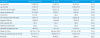
In KD 220 children, the mean age was 2.2±1.9 years, and the male to female ratio was 1.55:1. Most patients (n=191, 86.8%) had typical KD, and 29 (13.2%) had incomplete KD. Fifty-two patients (23.6%) were classified as having severe skin lesions, and 6 patients (3%) had arthritis. Total fever duration was 6.8±2.4 days, and 52 patients (23.6%) had CALs (Table 1).
2. Comparison of the patient groups with or without CALs
The group with CALs (n=52) was older (2.7±2.5 years vs. 2.1±1.6 years, P=0.031), and had longer fever duration (7.5±2.8 days vs. 6.6±2.2 days,P=0.040) and larger number of cases with repeated IVIG infusion (1.3 ±0.5 vs. 1.1±0.3, P=0.011), compared to the group without CALs (n=168). Patients with CALs had higher platelet counts than those without CALs (532±198 ×10 3 /µL vs. 465±160 ×10 3 /µL, P =0.013) and higher NLR values (2.7±3.8 vs. 1.7±1.4, P=0.004). Other clinical and laboratory findings were not significantly different between the two groups (Table 1).
3. Comparison of the patient gr oups with or without severe skin lesions
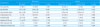
The patient group with severe skin lesions (n=52) had older age (3.1±2.3 years vs. 1.0±1.7 years, P<0.001), longer fever duration (P=0.041), and higher frequency of CALs (34.6% vs. 20.2%, P=0.033), compared to the group without severe skin lesions (n=168) (Table 2).
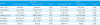
In laboratory parameters, patients with severe skin lesions had higher neutrophil differential (P=0.031), higher NLR (P=0.001), and more cases with pyuria (P= 0.001) (Table 3). The group with severe skin lesions tended to develop skin rashes earlier (4.1±1.8 days vs . 4.6±2.0 days, P=0.092) after disease onset, although not significantly, and had longer period of rashes after IVIG infusion (4.0±2.4 days vs. 2.7±1.9 days,P<0.001). A part of patients with severe skin rashes (36 cases) were prescribed antihistamines at 4.50±1.82 days after fever onset (not included in Tables).
4. Clinical characteristics of patients with arthritis
Among 6 patients with arthritis, 5 patients (83.3%) had oligoarticular lesions on the lower extremity joints, including the knee and ankle joints. The remaining 1 patient (16.7%) had polyarticular lesions on both multiple upper and lower extremity joints, including hands. Three (50%) of the 6 patients with arthritis were treated with MPD, and 2 patients (33.3%) were treated with infliximab because of IVIG non-responsiveness (Table 2). Although these parameters in the arthritis group showed a higher frequency than the patients without arthritis (n=214, P=0.004, respectively), the subjects in the former group was too small. There were no differences in clinical and laboratory parameters between the groups (Table 3).
Discussion
In the present study, we found that severity of skin rashes, one of clinical signs of KD, was associated with CALs. CALs, especially giant aneurysms, are a major complication and decide the prognosis in KD. It has been reported that clinical and laboratory parameters reflected the severity of systemic inflammation in KD, and they can help to predict the risk of CALs 512). The parameters include prolonged fever duration, IVIG non-responsiveness, and the elevated or decreased laboratory indices such as CRP, albumin, and sodium 5). In addition, a study reported that circulating plateletneutrophil aggregates might play a role in amplifying acute inflammation and CALs 14); another study found that NLR was higher in KD patients with CALs than those without CALs 15). In this series, we also found that the patients with CALs had significantly longer fever duration and higher platelet and NLR values, which may indicate more severe systemic inflammation.
In KD, skin rash usually appears 3–5 days after fever onset and presents as multiple symmetric, erythematous , papular, and macular exanthems. However, vesicles, crusted skin lesions, and itching sense are not usually evident. It is still unclear whether atypical skin lesions or severe cutaneous lesions are related to severe vasculitis, especially coronary artery inflammation. In addition, there were few studies regarding relationships between severe skin lesions and CALs. KD patients with atypical skin lesions such as pustular, vesicular or erythema multiforme like skin lesions were reported to have no CALs 1617). Whereas, KD patients with severe scattered crusting skin lesions or papules and keratotic lesions were correlated with CALs and refractory clinica l course 18).
In this series, KD patients with severe skin lesions such as generalized rash with desquamation and/or scattered crusting skin lesions with itching had longer fever duration and higher frequency of CALs. Severe skin lesions in KD tended to be observed earlier after fever onset and had significantly longer time to subside than mild skin rash. Also, patients with severe skin lesions tended to have higher neutrophil and platelet counts, and had significantly higher NLR values. Therefore, severe skin lesions in KD seems to be related to the severity of systemic inflammation and the risk of CALs.
The incidence of arthritis may be dramatically reduced in IVIG era as well as that of severe CALs (aneurysms) 1). Arthritis usually appears in the acute stage, but it can be observed after IVIG treatment 19). In articular types in KD, pauciarticular type was predominant to polyarticular type as well as in this study 120).
Recently, it has been reported that patients with systemic juvenile idiopathic arthritis (sJIA) in young children sometimes had similar clinical and immunological parameters with CALs 212223). These patients had more prolonged fever duration, treatment duration and higher age than typical KD patients.
In the present study, patients with arthritis had more additional treatment with IVIG, MPD, and infliximab, compared to those with other KD patients. These finding also suggest that severity of systemic inflammation is associated with CALs, regardless of phenotype of systemic immune-activation diseases.
The reasons of appearing CALs, skin rashes, and arthritis in KD remain to be answered. It is postulated that the main function of the host immune/repair system on the molecular level is to control the levels of toxic substances to the host cells; specific immune cells control distinct toxic protein substances based on their size and other characteristics. Host immune cells, including macrophage-lineage cells, control not only the pathogen-derived substances such as pathogen associated molecular patterns (PAMPs) but also the substances known as damage (danger)-associated molecular patterns (DAMPs) that may be derived from host cells injured by infectious insults 524). Since the substances that can bind to organ-specific cell receptors and corresponding immune cells to the substances may be responsible for inflammation in KD, it is possible that various clinical signs of KD are elicited by this mechanism. Thus, it could be explained that KD patients with giant aneurysms may have improper immune/repair system against the substances from KD agents and/or injured coronary artery cells 24).
This study had several limitations. Although we have been interested in skin lesions in KD, the selection of patients with severe skin rashes was done retrospectively with a subjective decision making. Another limitation was the small number of patients with arthritis, which made it impossible to compare CAL incidence. We believe that well-designed larger-scale studies are needed to obtain statistical significance.
In conclusion, our study showed that patients with severe clinical signs such as severe skin lesions or arthritis had severe systemic inflammation reflected by longer fever duration, some laboratory changes, and higher frequency of CALs. Pediatricians are needed to pay more attention to these patient groups for proper treatment on CAL prevention.
 XML Download