Dear Editor:
Erosive adenomatosis of the nipple (EAN) and mammary Paget's disease (MPD) are respectively a benign and a malignant neoplasm affecting the nipple/areola12. As the clinical characteristics of these conditions are usually nonspecific and variable, they are often mistaken for inflammatory skin diseases, such as psoriasis and eczema12. Recently, it has been shown that, in long-standing cases, dermoscopy may be used as an auxiliary tool in assisting their recognition12. However, there is no data on dermoscopy of such disorders in their initial stages, when the clinical identification is even more challenging. We here describe the dermoscopic features of two early instances of EAN and MPD.
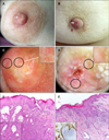
Case 1. A 41-year-old woman presented with a 1-month history of a subtle asymptomatic erosion associated with moderate hyperkeratosis on the right nipple (Fig. 1A). On noncontact polarized light dermoscopic examination, such lesion showed whitish/yellowish hyperkeratosis and sparse dotted vessels on a reddish-whitish background (Fig. 1C). Histology displayed typical features of EAN (Fig. 1E).
Case 2. An 88-year-old woman was referred to our clinic with a 1-month history of a small asymptomatic serohematic crust on the right nipple (Fig. 1B). Noncontact polarized light dermoscopy displayed a brownish-reddish crust associated with linear irregular vessels on a pinkish-whitish background (Fig. 1D). Histology and immunohistochemical analysis yielded a diagnosis of MPD (Fig. 1F).
The use of dermoscopy for assisting the diagnosis of skin conditions other than melanocytic lesions/common nonmelanocytic tumours has remarkably increased over the last years3. Regarding MPD, most of the attention has been focused on the pigmented variant, with diffuse irregular pigmentation, regression-like structures (especially gray-blue structures) and irregular vessels being described as the commonest findings by several reports45. According to a recent instance, some of the above-mentioned features may be found even in the classic (clinically nonpigmented) form of MPD, including light brown diffuse pigmentation, irregular black dots, irregularly distributed blue-gray dots (peppering), and irregular linear vessels; moreover, several chrysalis-like structures may additionally be detected1. On the other hand, based on a recent case report, dermoscopic examination of EAN may show cherry-red dots over a reddish background and nonspecific peripheral orange collar-like veils, interpreted by the authors as luminal openings and remaining epidermis, respectively2.
In the present two instances, we observed some of the findings reported in the two aforementioned long-standing cases of MPD and EAN, namely irregular linear vessels and sparse dotted red structures (which we interpreted as dotted vessels as they disappeared after pressing the dermoscope), respectively. Such a difference in dermoscopic vascular patterns might be explained by the well-known concept that benign lesions commonly present a regular vascularity and malignant lesions usually display more irregular vessels12. In our opinion, the detection of the above-mentioned findings might help the clinician raise the suspicion of such conditions (with consequent prompt biopsies) over the main differential diagnoses, i.e., eczema and psoriasis, as they usually show different features, namely yellowish serocrusts/patchily distributed dotted vessels and whitish scales/diffusely distributed dotted vessels, respectively3. Obviously, further studies on larger groups of patients are needed to confirm our observations.
Figures and Tables
Fig. 1
Clinical examination of the right nipple shows a slight erosion (arrow) associated with moderate hyperkeratosis in the first patient (A) and a small serohematic crust (arrow) in the second patient (B). Dermoscopic examination (carried out with DermLite DL3×10; 3Gen, San Juan Capistrano, CA, USA) of the right nipple in the first case displays whitish/yellowish hyperkeratosis and some dotted vessels (circles) on a reddish-whitish background (C), while it shows a brownish-reddish crust associated with linear irregular vessels (circles) on a pinkish-whitish background in the second case (D). Histologic examination in the first patient reveals typical features of erosive adenomatosis of the nipple, namely ductal structures in the dermis focally connected to the overlying epidermis (H&E, ×40); these ducts result mostly lined with a basal layer made up of myoepithelial cells and a luminal layer of epithelial cells exhibiting apocrine differentiation (box) (H&E, ×200) (E). In the second patient, histology shows large atypical cells with hyperchromatic eccentric nuclei and abundant cytoplasm throughout the epidermis, hyperkeratosis, parakeratosis, and acanthosis (H&E, ×200); immunohistochemical analysis demonstrates that the tumour cells are positive for cytokeratin 7 (box) (F). All these findings are consistent with the diagnosis of Paget's disease.

References
1. Takashima S, Fujita Y, Miyauchi T, Nomura T, Nishie W, Hamaoka H, et al. Dermoscopic observation in adenoma of the nipple. J Dermatol. 2015; 42:341–342.

2. Crignis GS, Abreu Ld, Buçard AM, Barcaui CB. Polarized dermoscopy of mammary Paget disease. An Bras Dermatol. 2013; 88:290–292.

3. Errichetti E, Stinco G. The practical usefulness of dermoscopy in general dermatology. G Ital Dermatol Venereol. 2015; 150:533–546.


 PDF
PDF ePub
ePub Citation
 Print
Print


 XML Download