

 PDF
PDF ePub
ePub Citation
 Print
Print


Dear Editor:
The presence of xanthomas can be a manifestation of various systemic disease. Primary biliary cirrhosis (PBC) and autoimmune hepatitis are autoimmune diseases that progressively destroy intrahepatic bile ducts and hepatocytes, respectively. The damage results in prolonged obstruction of the bile ducts, which leads to hypercholesterolaemia. Here, we present the case of a patient with PBC–autoimmune hepatitis overlap syndrome who presented with xanthoma striatum palmare.
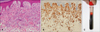
A 31-year-old female patient presented with multiple yellowish patches on the creases of both palms for 3 months (Fig. 1A). She had been diagnosed with PBC and concurrent autoimmune hepatitis 4 months ago. Her serum was positive for anti-mitochondrial, antinuclear, anti-smooth muscle, and anti-liver microsomal antibodies, with the latter three showing titers more than 1:80. A liver biopsy showed interface hepatitis consistent with autoimmune hepatitis, and chronic nonsuppurative destructive cholangitis of PBC. Serum biochemistry showed marked hypercholesterolemia, with high levels of total cholesterol (648 mg/dl) and low-density lipoprotein cholesterol (LDL-C, 495 mg/dl). Triglycerides (82 mg/dl) and high-density lipoprotein cholesterol (HDL-C, 72 mg/dl) were within normal ranges. Histopathologic examination revealed infiltration of the superficial dermis with numerous lipid-laden foamy histiocytes (Fig. 2A, B). A clear supernatant was noted when the patient's plasma was refrigerated at 4℃ for 24 hours, indicating type IIa hyperlipidemia according to Frederickson's classification (Fig. 2C). She was diagnosed with xanthoma striatum palmare, and treatment with a lipid-lowering agent (atorvastatin) was initiated. The cutaneous lesions on both palmar creases gradually flattened and faded out over 6 months (Fig. 1B). Follow-up laboratory tests of 6 months showed improved lipid profiles (LDL-C, 178 mg/dl; total cholesterol, 299 mg/dl; triglycerides, 94 mg/dl; HDL-C, 80 mg/dl).
The pathogenesis of xanthoma formation is explained by a complex process of dysregulation of macrophage cholesterol influx. Dysregulation of macrophage feedback leads to continuous cellular lipid accumulation, which finally results in the foamy cells seen on histological examination1. The distribution of xanthomas mainly on the palm is accounted for by the minor traumas to which these pressure-loading sites are exposed. Repeated minor trauma increases vascular permeability, with consequent leakage of lipids through the vascular endothelial wall. In PBC, prolonged obstruction of the biliary tree results in hypercholesterolemia, which over the longterm leads to xanthoma striatum palmare2.
PBC usually occurs alone, but in 7%~13% of cases, it may present as a syndrome in combination with autoimmune hepatitis3. The possibility of transition from autoimmune hepatitis to PBC has clinical significance, because the subsequent biliary tract obstruction may result in a xanthomatous condition3. To date, only three case of xanthoma striatum palmare has been reported regarding PBC245. No case of xanthoma striatum palmare in a patient with PBC–autoimmune hepatitis overlap syndrome has been reported so far. This case informs us that when a patient presents with yellowish plaque along palmar creases, an underlying hepatobiliary disease such as PBC and/or autoimmune hepatitis might be in the underlying related medical conditions.

 XML Download