

 PDF
PDF ePub
ePub Citation
 Print
Print


Dear Editor:
Acanthosis nigricans (AN) is characterized by hyperpigmented thickened skin with velvety texture in the flexural areas such as axillae, neck, groin, inner thigh, umbilicus, and perianal area. Obesity, insulin resistance, and hyperinsulinemia are often found in association with AN1. Vitiligo is an autoimmune disease which shows decreased melanin and melanocyte in epidermis. It is associated with autoimmune diseases such as thyroid disease and diabetes mellitus1.
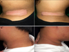
A 16-year-old male patient presented with hypopigmented mildly erythematous patch within hyperpigmented patch on neck and shoulder (Fig. 1). The preceding hyperpigmented patch appeared 6 years ago when the patient was treating his obesity and impaired insulin tolerance. The hypopigmented lesion appeared 6 months prior to the visit and gradually grew in size. The patient did not remember any history of trauma before the lesion appeared. Although the patient had previously taken oral metformin for a year, he was not taking any medication at the time of visit. No similar skin lesions could be found in family members of the patient.
Skin biopsy was performed on both hyperpigmented and hypopigmented lesions. In the former, typical features of AN such as hyperkeratosis, papillomatosis, and basal layer hyperpigmentation were observed. In the latter, however, S-100 protein stain and Fontana-Masson stain did not reveal remarkable decrease in both epidermal melanocytes and degree of basal pigmentation (Fig. 2).
The patient applied topical steroid and tretinoin for a year, resulting in complete repigmentation (Fig. 1). In contrast, the patient has kept his body weight and no significant change was seen in the AN lesion.
Up to this day there are four reports of AN accompanied by vitiligo1234. Out of these, histologic confirmation of depigmented epidermis was conducted in two cases12. Harman et al.1 reported a case of AN with partial depigmentation which developed with obesity. The AN lesion disappeared after substantial weight loss but the vitiligo lesion persisted. Garzitto et al.2 reported a case of malignant AN in a patient with preexisting vitiligo. Though the ovarian malignancy was surgically resected, both AN and vitiligo persisted.
This case is unique in two aspects. First, no significant loss of melanocyte and melanin pigment was observed. A traditional histologic feature of vitiligo is epidermal loss of melanocytes, but a recent research by Kim et al.5 imply that residual melanocytes, although in lower level, may exist in vitiligo lesion. Unfortunately the relationship between the amount of residual melanocyte and degree of repigmentation is not yet established. Secondly, the vitiligo lesion showed complete pigmentation after treatment. Vitiligo itself is not an incurable disease, but all the reports of vitiligo with concomitant AN have failed to induce repigmentation.
In conclusion, when AN is accompanied by hypopigmentation assessing the amount of melanocyte by means of skin biopsy can be helpful in predicting the potential degree of repigmentation after vitiligo treatment. This finding does not seem to affect the course of AN and thus treatment of underlying cause of AN would still be required.

 XML Download