Dear Editor:
Previously, we discovered the presence of specialized nail mesenchyme containing onychofibroblasts in the nail units which were obtained from polydactyly operations and proposed a new concept of onychodermis for the specialized nail mesenchyme1234. Furthermore, the presence of onychodermis was also demonstrated by CD10 immunohistochemistry in developing nail organ and small biopsy specimens from the adult nail unit56. However, very recently, it was reported that the concept of onychodermis was not applicable in normal adult nail unit7.
Thus, we performed this investigation to show obviously the presence and localization of onychodermis in normal adult nail unit.
Normal nail units were obtained from cadavers of four adults who had consented to scientific research. The nail units were longitudinally or transversely sectioned. They were fixed in formalin solution for paraffin blocks. Hematoxylin and eosin (H&E) staining was performed. Immunohistochemistry was applied to the sections, using a monoclonal antibody against CD10 (1:50; Clone 56C6; Novocastra, Newcastle, UK).
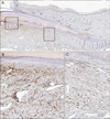
In H&E staining, the onychodermis below the nail matrix and nail bed was less eosinophilic, compared with the dermis of the other parts (Fig. 1). The presence of onychodermis was less definitely observed in normal adult nail unit than that in previously reported polydactyly samples3. However, CD10 was distinctly expressed in the onychodermis below the nail matrix and nail bed, while CD10 was negative in the dermis of the other parts of the nail unit (Fig. 2). Based on the results of histology and CD10 immunohistochemistry, onychodermis was very close to the nail bed and the distal part of the nail matrix, but it stood a bit aside from the proximal part of the nail matrix. In all cases we found very similar results about the presence and localization of onychodermis.
In this study, the selective expression of CD10 in the mesenchyme below the nail matrix and nail bed of normal adult nail unit, which was consistent with the finding in the nail unit which was obtained from polydactyly operation, supports the presence of the onychodermis (specialized nail mesenchyme) in normal adult nail unit as well. In the very recent paper, Perrin reported that there was loose connective tissue below the nail matrix, which was located a bit aside from the nail matrix7. Although he did not show the data of CD10 immunohistochemistry in that area, based on our study, the area of the loose connective tissue was CD10 positive and thus it belongs to the onychodermis.
CD10 expression in the onychodermis of the nail unit might be reasonable because it was expressed in periadnexal mesenchyme of the skin, such as the dermal sheath of hair follicle8 and dermal papilla and sheath of hair follicles in nevus sebaceous (data not shown). In our previous study, using the nail unit of the infant (polydactyly samples), there was a mesenchymal area called onychodermis, which showed much more cellularity and less eosinophilic, loose connective tissue beneath the nail matrix and the nail bed3. However, in this study, using the normal adult nail unit, the presence of onychodermis was less marked compared with that of the nail unit of an infant. The slight morphologic difference of the onychodermis between the adult and infant might be the change according to aging.
In the previous in vitro study using skin equivalent models, hard keratin expression was induced in the non-nail-matrical keratinocytes by the fibroblasts around the nail matrix through the epithelial–mesenchymal interactions9. Based on this finding and our results, the specialized nail mesenchyme (onychodermis) containing onychofibroblasts may play an important role in nail formation and growth3. One of problems in nail research is the difficulty in obtaining high-quality sections of the nail unit, especially in adult nail tissue because nails are extremely hard and tend to split or tear. Also, treatment of decalcification agents for sectioning adult nail tissue including hard phalangeal bone may affect the immunohistochemical staining, possibly resulting in weaker immunoreactivity.
Taken together, we confirmed that the concept of onychodermis (specialized nail mesenchyme) is applicable in normal adult nail unit.
Recently, we suggested that onychodermis might be involved in the development of onychomatricoma based on histology and CD10 immunohistochemistry10. Further investigation such as the function of the onychodermis and the relation of the onychodermis to the nail diseases will be necessary.
Figures and Tables
References
1. Lee KJ, Kim WS, Lee JH, Yang JM, Lee ES, Mun GH, et al. CD10, a marker for specialized mesenchymal cells (onychofibroblasts) in the nail unit. J Dermatol Sci. 2006; 42:65–67.

2. Lee DY, Yang JM, Mun GH, Jang KT, Cho KH. Immunohistochemical study of specialized nail mesenchyme containing onychofibroblasts in transverse sections of the nail unit. Am J Dermatopathol. 2011; 33:266–270.

3. Lee DY, Park JH, Shin HT, Yang JM, Jang KT, Kwon GY, et al. The presence and localization of onychodermis (specialized nail mesenchyme) containing onychofibroblasts in the nail unit: a morphological and immunohistochemical study. Histopathology. 2012; 61:123–130.

4. Lee DY, Park JH, Shin HT, Jang KT, Kwon GY, Shim JS. Onychodermis (specialized nail mesenchyme) containing onychofibroblasts in horizontal sections of the nail unit. Br J Dermatol. 2012; 166:1127–1129.

5. Sellheyer K, Nelson P. The concept of the onychodermis (specialized nail mesenchyme): an embryological assessment and a comparative analysis with the hair follicle. J Cutan Pathol. 2013; 40:463–471.

6. Park JH, Lee DY. CD10 is expressed in nail mesenchyme (onychodermis) containing onychofibroblasts of the adult nail unit. Clin Exp Dermatol. 2012; 37:685–686.

7. Perrin C. The nail dermis: from microanatomy to constitutive modelling. Histopathology. 2015; 66:864–872.

8. Lee KJ, Choi YL, Kim WS, Lee JH, Yang JM, Lee ES, et al. CD10 is expressed in dermal sheath cells of the hair follicles in human scalp. Br J Dermatol. 2006; 155:858–860.


 PDF
PDF ePub
ePub Citation
 Print
Print


 XML Download