

 PDF
PDF ePub
ePub Citation
 Print
Print


INTRODUCTION
Burn wound healing is a complex process that involves various mediators and multiple cell types around the injured zone. A second-degree burn, also called a partial thickness burn, causes the skin to blisters. Burn blisters fluid might be important roles in wound healing process1. Some reports indicate that retention fluid from blister of partial thickness burn, which contains relatively large amounts of cytokines and soluble mediator, may affect the wound healing process234. However, the effect of burn blister fluid on wound healing has not been fully explored.
Heat shock protein 70 (HSP70, molecular weight 70 kDa) is the most well understood member of the heat shock family of proteins. HSP is found in all cells and has a chaperone function during the assembly process of protein folding and polypeptide formation as well as a cell protection function. Specifically, extracellular HSP70 is involved in intercellular signaling and is involved in the innate, adaptive immunologic response, interrupts apoptosis, and protects cell structure as a chaperone567. HSP70 can both induce and arrest inflammatory reactions in experimental brain injury and ischemia. It is reported that HSP70 is involved in the healing process, as it has been found in the epidermis and dermis of thermal injury patients8910. If HSP70 is present in burn blisters, it likely affects the damaged cells of thermal injury patients.
Inflammation is an integral phase of wound healing. The local inflammatory response of a skin wound is characterized by increased amounts of several pro-inflammatory cytokines, such as interleukin-8 (IL-8), tumor necrosis factor-α, and prostaglandin E2, and local neutrophil infiltration11. Excessive and ongoing wound inflammation inhibits the healing of burn wounds by fostering an unabated influx of leukocytes to the wound site12, which directly contributes to the progressive deepening and extension of the wound13. This inflammatory response is amplified by an elevated IL-8 level in the wound tissue14. However, IL-8 has also been shown to act on epithelial cells or endothelial cells to promote migration, invasion, proliferation, and angiogenesis in vivo and enhanced wound healing due to induced reepithelialization151617.
There are various opinions regarding the treatment method for the blisters of partial thickness burns, including removing the blister roof, extracting the fluid maintaining the blister roof, or leaving the blister intact181920. A better understanding of the changes in heat shock proteins and pro-inflammatory factors over time is expected to lead to an improved blister management strategy.
This study aimed to determine whether HSP70 and IL-8 are present in burn blisters, the variation in their concentrations over time, and the influence of HSP70 concentration on the length of the treatment period.
MATERIALS AND METHODS
Study subjects
For this study, patients with partial thickness burn with bullae who were aged 18 years and over and agreed to cooperate during the period from October 1, 2013 to March 31, 2014 were selected. Patients with immune-related diseases, with diseases that can induce an immune unbalance, and who were undergoing cancer treatment or other hormonal treatments were excluded. For all patients, information regarding sex, age, treatment period, total burn surface area (TBSA), and the abbreviated burn severity index (ABSI) score was obtained. The protocol for this study was reviewed and approved by the institutional Ethics Committee for human studies of Hangang Sacred Heart Hospital, Burn Center, Seoul, Korea (2013-068). We considered p-values<0.05 to be statistically significant.
Collection of blister fluid
After informed consent was obtained, blister fluid was aspirated from the intact burn blisters of patients with thermal burns who were admitted to our Emergency Department of Hangang Sacred Heart Hospital. Aspiration of the blister fluid was performed with an 18-gauge needle. The bullous fluid was first collected while leaving the blister roof intact. On the next visit, the fluid from the same bullae was collected if fluid remained. The collected amount for each case was 1 ml. We set the time after injury to sampling as the collected time. The first day was defined as the time between 1 hour and 12 hours after the damage occurred, the second day was between 25 and 36 hours, the third day was between 49 and 60 hours, the fourth day was between 73 and 84 hours, and the fifth day was between 97 and 108 hours. To ensure consistent conditions, any case that did not meet the criterion was excluded from the analysis. The end of the treatment period was determined by a plastic surgeon according to the treatment results.
Heat shock protein 70 and interleukin-8 quantification in blister fluid
The concentrations of HSP70 and IL-8 were measured using enzyme-linked immunosorbent assay (ELISA) kits. ELISA was used to measure IL-8 and HSP70 in the bullae of burn patients. The quantitative measurement of IL-8 and HSP70 was performed in duplicate samples using commercial ELISA kits (Cat. No. KAC1301; BioSource, Nivelles, Belgium) for IL-8 and HSP70 (Cat. No. ab13306; Abcam, Cambridge, UK) according to the manufacturer's instructions. Each sample was diluted 100-fold (HSP70) or 5-fold (IL-8) with dilution solution, and the samples were centrifuged for 5 minutes at 4℃ and 12,000 rpm to remove other debris and blood cell components. Then, 100 µl of the standard and bullae samples were placed in an antigen-coated 96-well plate and left for 2 hours at room temperature, after which they were washed four times with 400 µl washing solution. Next, anti-HSP70 and anti-IL-8 conjugates were placed in the wells followed by incubation for 2 hours at room temperature. After washing, each well was filled with a freshly prepared chromogenic solution, and the plate was incubated for 30 minutes. Finally, the reaction was stopped with by adding stop solution (1.8 N H2SO4), and the plate was read at 450 nm using a DTX880 multimode detector (Beckman Coulter, Brea, CA, USA).
Statistical analysis
All statistical analyses were conducted using PASW Statistics 18.0 (IBM Co., Armonk, NY, USA). The results were expressed as means±standard deviations or median values and interquartile range (IQR). A correlation analysis was performed to determine the relationship between the concentration of HSP70 and IL-8 and the length of the treatment period. The correlation analysis was performed using the Spearman test. We used the Mann-Whitney U-test to compare non-normally distributed variables.
RESULTS
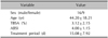
Among 53 total cases (29 patients), 36 cases (25 patients) that met the inclusion criteria were analyzed. Twenty cases from the first day, 6 cases from the second day, 5 cases from the third day, 3 cases from the fourth day, and 2 cases from the fifth day were evaluated. Table 1 presents the demographic data of the patients. Sixteen patients were male, and 9 patients were female. The average age was 44.20 years old (minimum, 18 years old; maximum, 90 years old), the average TBSA was 3.12% (minimum, 1%; maximum, 8%), and the average ABSI score was 4.00 (minimum, 2; maximum, 7). The average treatment period was 15.08 days (minimum, 6 days; maximum, 37 days). Age, TBSA, ABSI score, and treatment period were not normally distributed, and there was no significant correlation between them and HSP70 and IL-8.
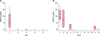
Heat shock protein 70
The median concentration of HSP70 was the highest on the first day (median, 5.555 ng/dl; IQR, 1.602~12.164 ng/dl). On the second day and third day, the median values were 0.038 ng/dl (IQR, 0.007~0.058 ng/dl) and 0.170 ng/dl (IQR, 0.058~0.552 ng/dl), respectively. HSP70 was not detected on the fourth and the fifth days (Fig. 1A). The majority of the HSP70 was found in the first 12 hours. The highest median HSP70 value was measured 1 hour after the damage, at 9.261 ng/dl (IQR, 7.347~15.345 ng/dl). After 1 hour, the concentration decreased rapidly: 7.285 ng/dl (IQR, 1.732~14.515 ng/dl) after 2 hours, 2.038 ng/dl after 4 hours, 0 ng/dl after 5 hours, 1.264 ng/dl after 11 hours, and 0.182 ng/dl after 12 hours (Fig. 1B). There was no correlation between the concentration of HSP70 and treatment period during the first 12 hours (rho=−0.316, p=0.175).
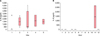
Interleukin-8
The concentration of IL-8 was measured from the first day to the fifth day. The median concentration of IL-8 was 0 pg/dl on the first day, 1,477.245 pg/dl (IQR, 733.622~2,302.500 pg/dl) on the second day, 511.429 pg/dl (IQR, 322.483~4,021.122 pg/dl) on the third day, 1,653.010 pg/dl (IQR, 584.388~2,187.322 pg/dl) on the fourth day, and 1,473.6735 pg/dl (IQR, 1,006.071~1,707.477 pg/dl) on the fifth day (Fig. 2A). When the concentration of IL-8 within the first 12 hours was analyzed, IL-8 was not detected up to 5 hours after the damage (Fig. 2B). There was no correlation between the concentration of IL-8 and treatment period (rho=−0.326, p=0.053).
DISCUSSION
HSP70 is released in a passive manner in necrotic cells and is also secreted through an active mechanism involving lysosomal vesicles and lipid rafts21. The functions of HSP70 include acting as a molecular chaperone to facilitate the assembly of proteins, targeting misfolded and damaged proteins for degradation, dissembling protein aggregates, acting as a regulatory molecule in protecting cells from thermal or oxidative proteins, and inhibiting apoptosis8. In addition, HSP70 is involved in inflammatory reactions and the healing process of the epidermis and dermis following thermal injury8910. HSP70 has an extracellular function that appears to potentiate the immune response, whereas the intracellular mechanisms appear to be anti-inflammatory. HSP70 can also interrupt the cleavage of pro-matrix metalloproteinases to an active form, which in turn may reduce immune signaling22.
The concentration of HSP70 was found to be very high only on the first day. It was weakly detected on the second day. A small amount was found on the third day. Finally, no detectable amount was found on the fourth and the fifth days. Because the concentration of HSP70 was the highest in the first 12 hours, the results from 1 hour to 12 hours after the burn injury were analyzed in detail. The concentration was the highest 1 hour after the injury (Fig. 1B). Then, the concentration decreased rapidly afterwards. It was weakly detected after 5 hours. Even though there were not many cases included in this study, the results indicated a pattern that HSP70 is present in blisters for the first few hours, after which the amount decreases. In this study, no correlation was found between the concentration of HSP70 during the first 12 hours and the length of the treatment period (rho=−0.316, p=0.175).
A major burn injury induces an inflammatory response, which is accompanied by the release of various cytokines23. The chemokine IL-8 induces chemotaxis in neutrophil granulocytes (polymorphonuclear leukocytes, PMNs), causing them to adhere to endothelial cells and then move into tissue cells to induce PMN-mediated damage24. Therefore, many inflammatory mediators are involved in multiple steps of the systemic inflammatory response syndrome to a trauma or severe burn, and they may be used as an index to estimate the extent of the inflammatory response or prognosis. IL-8 has been shown to play a pivotal role in the activation of the pro-inflammatory cascade in necrotizing enterocolitis (NEC)2526, and it has been recently demonstrated that IL-8 may serve as a potential predictive marker in diagnosing NEC. Hans-Oliver reported that IL-8 increases the amount of the intracellular integrin α6 subunit, keratinocytes proliferation, and the number of mitotic keratinocytes and enhances reepithelialization17. Berney et al.27 reported that the serum IL-8 concentration increased after several days or the first day in acute pancreatitis, reaching a plateau between days 2 and 5, and also reported that the concentration of serum IL-8 in a severe case increased on the day the patient visited the emergency department. Lee et al.28 reported that immunomodulatory effect of interleukine expressed.
In this study, the changes in IL-8 levels in burn blister fluid were investigated. The IL-8 concentration in partial thickness burn cases was found to be the highest on the fourth day. IL-8 was not detected up to five hours, but it was detected after 11 hours. Cases for 6 to 10 hours are not included in this study. However, no relationship between the IL-8 concentration in burn blisters and the length of the treatment period was found.
Our results suggest that IL-8 is an important mediator in inflammatory changes following a burn injury but that various factors may influence the concentrations of this cytokine. Luo et al.29 demonstrated a novel role for exogenous human HSP70 in suppressing the production of IL-6, IL-8, and monocyte chemoattractant protein-1 in fibroblast-like synoviocytes that involves inhibiting the activation of the mitogen-activated protein kinases and nuclear factor-κB signaling pathways.
There is still a debate regarding how to best treat the blisters of burn patients192021. This study showed that HSP70 increased in the first few hours and decreased afterwards and that IL-8 was produced after a few hours and increased afterwards. It is speculated that HSP70 present in a blister has a pro-inflammatory effect outside the cell and a protective effect in the intracellular space of the ischemic cell zone30. If this is the case, then the proper treatment would be to remove the blister fluid during the period when HSP70 is present. In addition, a better understanding of the changes in IL-8 concentration in partial thickness burn blisters would help to improve blister management.
Because this study was based on patient schedules, it was not possible to obtain all of the data on the cases for the targeted times. In addition, the number of cases was limited because we counted the first 12 hours as one day; thus, we likely lost some cases, and there were many cases for which we could not re-sample the fluid from the same blister.
This study had limitations and could not clearly define the healing mechanism in burn blister fluid, but the results demonstrate the changes in the concentrations of HSP70 and IL-8 and may help determine the guidelines for when blister fluid should be removed.
Various inflammatory mediators function during the healing process. To better understand the immunological processes during wound healing and how to best treat wounds, additional studies on the changes in HSP70 and IL-8 levels over time as well as those of other mediators are necessary.
 XML Download