

 PDF
PDF ePub
ePub Citation
 Print
Print


INTRODUCTION
Reducing facial wrinkles has been one of the more popular cosmetic aims, as with people wanting to live healthier, they also want to look younger. Consequently, the use of anti-wrinkle treatments and functional cosmetics is increasing1. For example, anti-wrinkle treatments such as laser therapy, various dermal filler injections and invasive procedures like thread lifting are becoming more widely used23. However, such treatments are usually expensive, they may cause discomfort, and are procedures that require professional intervention.
Skin wrinkles appear due to loss of elasticity caused by rapid degradation of collagen4. These changes are influenced by intrinsic and extrinsic factors that involve mitogenic reactions and signal transduction pathways. Many receptors for epidermal growth factor (EGF), platelet-derived growth factor, interleukin-1, tumor necrosis factor participate in this process5. EGF stimulates and regulates the proliferation of various cell types in vitro6, and for skin cell types, EGF plays an important role in growth and regeneration of keratinocytes and fibroblasts. As such, EGF might be a potential therapeutic and cosmetic agent for damaged skin and injuries including wrinkles and aging7.
The epidermis of skin contains stratum corneum, which consists of keratinocytes that act as “bricks” and intercellular lipids that act as “mortar.” Intracellular lipids act as a barrier against many agents including drugs and various cosmetic agents891011. For these reasons, a topical application of EGF has limited efficacy on treating wrinkles.
Various studies have been conducted to find ways to improve the skin penetration of drugs and cosmetics. For example, micro-needles for transdermal drug delivery have been developed in various forms1213. Micro-needle patches have also been recently used to increase skin permeability, improving drug delivery and for also for various cosmetic purposes14. However this technology often allows for insufficient skin penetration, and there is also low compliance with its use due to discomfort. Accordingly, we developed micro-spicules containing epidermal growth factor (MS-EGF, also called micro-needle EGF) to increase dermal penetration of EGF, and in this study, we investigated the feasibility of MS-EGF as a new technique for drug delivery by assessing its efficacy on wrinkle reduction, along with its safety profile.
MATERIALS AND METHODS
Penetration assay for micro-spicule containing epidermal growth factor
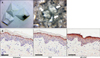
MS-EGF is a 0.25-µm pyramidal shaped material composed of hyaluronic acid. This micro-spicule contains EGF and is readily soluble in water. MS-EGF cream is MS-EGF mixed with a general hydrophobic cosmetic paste (Fig. 1A).
We performed an ex vivo skin penetration test using pig skin (Medikinetics, Pyeongtaek, Korea) to investigate the penetration ability of MS-EGF in epidermis. The pig skin samples were divided into three groups: untreated, MS-EGF and EGF alone group. For the MS-EGF group, 100 mg of MS-EGF was scrubbed for 20 seconds and then distilled water was added to dissolve the remaining micro-spicules, followed by additional rubbing for 20 seconds. For the EGF alone group, 100 mg of EGF was applied for 40 seconds. All the sample groups were observed at room temperature for 10 minutes and then stored at 4℃ for 12 hours. Tissue samples were fixed with 10% formaldehyde, embedded in paraffin, and cut into sections. The sections were deparaffinized and processed with Bond™ polymer refine detection kit (Leica Microsystems, Wetzlar, Germany). The primary EGF antibody (Santa Cruz Biotechnology, Dallas, TX, USA) was diluted to 1:50 and the sections were incubated with secondary antibody at room temperature for 10 minutes. The sections were incubated with diaminobenzidine tetrachloride solution at room temperature for 5 minutes and counterstained with 0.1% Mayer's hematoxylin.
Subjects and study design
Twenty healthy volunteers were enrolled in this randomized, controlled, left side-right side split-face test. The participants were over 30 years old with mild to moderate periocular wrinkles. All the volunteers were exposed to the same external environment. Demographic data such as age, gender, and past medical history was collected prior to enrollment. Volunteers receiving therapeutic interventions such as botulinum toxin or fillers and those with concomitant cutaneous diseases such as acne and scar on periocular region were excluded from the study.
Participants were asked to apply either MS-EGF cream or EGF cream on each periocular wrinkle on a daily basis for 4 weeks. MS-EGF participants were told to apply the cream for 20 seconds, add distilled water to dissolve the remaining micro-spicules for the purpose of preventing irritation, and then rubbing again for 20 seconds. EGF alone participants were told to apply the cream for 40 seconds with the same intensity. This study was approved by the Institutional Review Board of Chungnam National University Hospital (CNUH 2015-05-013). All of the subjects provided written informed consents before participating in the study.
Clinical outcome evaluation
The participants were assessed at the beginning of the treatment and at 1, 2, 4, and 8 weeks of treatment regimen. A-One Pro digital skin image analysis equipment (Bomtech Electronics Co., Seoul, Korea) for objective evaluation of wrinkle degree and 3-dimensional (3D) skin image of periocular wrinkles was used during each visit. Dermal density and dermal depth were measured by ultrasonic equipment, Ultrascan UC22 (Courage Khazaka Electronics, Cologne, Germany). The severity of periocular wrinkles was assessed according to a 5-point photonumeric scale. The scale ratings are: 0 for no wrinkles, 1 for very fine wrinkles, 2 for fine wrinkles, 3 for moderate wrinkles, and 4 for severe wrinkles15. At the end of the study, the patients documented their degree of satisfaction as very satisfied, satisfied, slightly satisfied, or unsatisfied. Patients were also asked to report any side effects during each visit.
Statistical analysis
Statistical analyses were performed using SPSS ver. 15.0 (SSPS Inc., Chicago, IL, USA). The Mann-Whitney U-test was used for post hoc analysis. To compare the categorical data, the chi-square test or the Fisher exact test was performed. Correlations were performed using the Spearman correlation analysis. p-values<0.05 were considered to be statistically significant.
RESULTS
In a preliminary study using pig skin, stratum corneum was effectively removed and EGF in the epidermis was uniformly distributed in the MS-EGF group. EGF alone group also showed a weak presence of EGF in the epidermis; however, there was no significant difference compared to the untreated group (Fig. 1B).
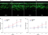
A randomized, controlled, left-right face spilt test was performed afterwards to test the efficacy of MS-EGF on periocular wrinkle. A total of 20 healthy volunteers (16 females, 4 males) with an average age of 45.2 years (range, 33~54 years) were enrolled in this study. There were no significant differences in the degree of baseline wrinkles between each group. After daily applications for each group for 4 weeks on each periocular wrinkle, the MS-EGF grouped showed a marked improvement at 8 weeks after topical application, shown with skin ultrasound analysis (Fig. 2A). The mean dermal density of both groups at baseline was similar (6.25%±1.73% in MS-EGF vs. 6.38%±1.55% in EGF alone) and this value was set as a standard of the dermal density ratio changes. In the MS-EGF group, dermal density ratio was significantly improved at 4 and 8 weeks after treatment, respectively (p<.0.01, p<.0.001). In the EGF alone group, there was a slight increase in dermal density, but the difference was not statistically significant. The MS-EGF group also demonstrated a significant increase in dermal density after 4 and 8 weeks compared to EGF alone group, respectively (p<.0.05, p<.0.01; Fig. 2B).
Results for mean dermal depth were similar to those for dermal density. Although mean dermal depths of both groups at baseline were similar (1.75±0.30 mm in MS-EGF vs. 1.80±0.46 mm in EGF alone), MS-EGF group showed a significantly increase in dermal depth after 8 weeks compared to the EGF alone group (p<.0.05; Fig. 2C). From the above observations, there was a 30.1% increase in dermal density and a 19.5% increase in dermal depth from baseline in the MS-EGF group.
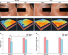
Clinical digital photographs and 3D image of periocular wrinkles by A-One Pro image analysis were used for clinical comparisons. Although both groups showed favorable results after 8 weeks compared to the baseline, the clinical photograph and 3D skin image improved significantly more with MS-EGF treatment than that for EGF alone treatment (Fig. 3A, B). Wrinkle severity score was evaluated automatically with A-One Pro, using a 5-point photonumeric scale. In the A-One Pro measurements, wrinkle scores decreased in both groups; however, the MS-EGF group showed significant improvement in wrinkle score compared to EGF alone group after 8 weeks (p<.0.05) (Fig. 3C). In the 5-point photonumeric scale, there was no significant difference in the grades between the two groups; however, the MS-EGF group showed a greater change in mean grade using this scale (2.5~2.0 in MS-EGF vs. 2.4~2.2 in EGF alone group; Fig. 3D).
Subjective improvement was also assessed by all participants upon completing the study. Among the participants who received MS-EGF treatment, 70% were “very satisfied” or “satisfied” after the treatment, whereas only 15% were “dissatisfied.” For the EGF alone group, 50% were “very satisfied” or “satisfied” after treatment whereas 20% were “dissatisfied” (Fig. 4). The treatments were well-tolerated by most patients and there were no noticeable adverse events such as inflammation, desquamation and pigmentation in either of the treated areas. Six participants in MS-EGF experienced transient erythema and a prickling sensation immediately after the application, which resolved within a few minutes without special management.
DISCUSSION
Wrinkles are one of the most common morphological features of skin aging16. Interest in anti-aging is growing every day as people pay more attention to beauty and their looks. For these reasons, the interest in functional anti-aging cosmetics has increased for most age groups117, and many cosmetic products targeted at reducing signs of aging have been developed. Despite various approaches in developing cosmetics, these products have limited skin penetration due to the skin barrier. Therefore, various molecular, chemical, and morphological studies have been performed on increasing the skin penetration of various topical agents18192021.
Micro-needle technology offers an efficient and minimally invasive drug delivery compared to conventional transdermal patches and intradermal injections22. With the advancement of micro-units manufacturing technology, micro-needles have been developed by academic laboratories and pharmaceutical companies, and are currently being used to enhance transdermal delivery of various molecules2324. Micro-needles increase the patients' compliance as they may have a regular needle phobia so that patients can apply the drug by themselves25. Micro-needles in form of patches are band-aid like materials with embedded micro-needles fabricated in arrays and can be in four versions such as hollow, solid, coated and polymer forms. However, there are some limitations for using these patches. First, skin pore closure needs to be additionally investigated after micro-needle patches application as it relates to the risk of infections. Second, it is difficult to control the drug amount in micro-needle patches26. To overcome these challenges, a new drug delivery technique with higher efficacy and safety is required.
This was the first study to investigate a soluble micro-spicule type topical agent for anti-wrinkle improvement. In experiments using pig skin, micro-spicule cream showed higher skin penetration ability than a typical cream, containing the same ingredients. This method allows delivery of various agents, but with a topical application. As such, it was possible to evaluate the anti-wrinkle efficacy and safety of soluble MS-EGF, delivering EGF beyond the external skin barrier.
In the randomized and controlled face spilt trial of 20 healthy volunteers who had MS-EGF and EGF in a cream form, periocular wrinkles were clinically improved in both groups after 8 weeks. However, there were significant differences in dermal density, dermal depth, and wrinkle severity after 4 and 8 weeks between the two treatment forms. Anti-wrinkle improvement with MS-EGF was more significant and superior to the EGF alone treatment. In the study, the skin penetration ability for EGF was also larger with MS-EGF. This implied the increased anti-wrinkle efficacy of MS-EGF was due to superior skin penetration ability from the micro-spicules. Most participants were more satisfied when using MS-EGF and consider that micro-spicules might find more applications and be more favored, despite the scrubbing of particles, and regardless of pain during the application. Although there was a temporary erythema and mild pain on MS-EGF lesion when rubbing strongly, it led to a better clinical outcome.
There has been one report on clinical improvements of periocular wrinkle using micro-needle patch including EGF. It showed the positive effects for the micro-needle EGF patch on physician-rated wrinkle scores, patient satisfaction levels, and corneometer results. However, there were no statistical differences compared with the control group, hyaluronic acid-based and without EGF micro-needle patches. This implied that the anti-wrinkle effects of the micro-needle patch may had been solely be due to the HA rather than EGF in the patches27. To further delineate the advantage of MS-EGF, in the future, additional studies need to compare the efficacy of EGF micro-needle patches with MS-EGF cream technology for wrinkle reduction.
In conclusion, the results of this study identified a favorable clinical benefit for MS-EGF in treating periocular wrinkles along with good tolerability and higher satisfaction from the volunteers that used it. Furthermore, the soluble property of micro-spicules is noteworthy for a new type of cosmetics delivery system and has more development potential in various other applications.

 XML Download