

 PDF
PDF ePub
ePub Citation
 Print
Print


INTRODUCTION
Phytophotodermatitis is a condition caused by sequential exposure to certain photosensitizing substance present in plants followed by sunlight. Many common plants, including citrus fruits, celery, and wild parsnip contain such photosensitizers (e.g., furocoumarins).
Herein, we present five cases of phytophotodermatitis after soaking feet in fig leaves decoction.
We present two purposes of this report: the first is to bring attention to this type of dermatitis. It is important for dermatologists to recognize phytophotodermatitis as it may sometimes be misdiagnosed as other skin conditions including cellulitis, tinea, and allergic contact dermatitis. The cases described here show fig leaves-induced phytophotodermatitis. The second purpose is to suggest that the use of fig leaves as folk remedy not only has no scientific basis but may also cause severe adverse events.
CASE REPORT
Case 1
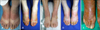
A 57-year-old female patient presented an erythematous swollen patch with bullae on both hands and feet for three days after using traditional herbal medicines (Fig. 1A).
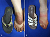
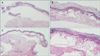
She applied fig leaves decoction for psoriatic lesion on both feet. She received the treatment for six or seven times during a week. Afterward, she walked outside on a sunny day. Five hours after the exposure to the sun (ultraviolet light), she felt a burning sensation in both hands and feet, and an erythema had developed on the feet dorsa; the lesions were hidden under the straps of the sandal (Fig. 2A). There was no history of a similar rash in the past or any form of drug or medication use. She was otherwise systemically well and did not have any personal or family history of photosensitivity. Histopathologic examination revealed sub-epithelial blisters with intensive epidermal necrosis (Fig. 3A, B).
On follow-up examination, after a 4-month period using systemic and topical steroids, improvements were observed.
Case 2
A 69-year-old woman presented with erythematous patches with bullae on the photo-exposed area of both feet, 7 hours after exposure to fig leaves decoction and sunlight (Fig. 1B, Fig. 2B). A detailed history revealed that she had applied a self-prepared fig leaf remedy to treat her onychomycosis. She had been soaking her feet in the decoction water three times daily for two days. Skin biopsy showed full epidermal necrolysis (Fig. 3C, D). Treatment included the administration of systemic steroids, antihistamines, and the application of topical antibacterial creams. Her lesions resolved after two months.
Case 3
A 66-year-old man developed a sharply defined erythematous swollen patch with bullae on both feet (Fig. 1C). The previous day, he had soaked his feet in fig leaves decoction to treat his onychomycosis. Afterwards, he walked outside on sunny day. His lesions were treated with systemic and topical steroids, but he was not available for skin biopsy and follow-up.
Case 4
An 87-year-old man presented with extensive skin lesions. On examination, erythema, edema, vesicles, and bullae were observed exclusively on both feet (Fig. 1D). When questioned about the history, he remembered that the previous day he had soaked his feet in fig leaves decoction to treat his onychomycosis. He soaked his feet three times a day and spent time in the outside. The patient refused skin biopsy. He was treated with systemic and topical steroids with instructions to avoid exposure to sunlight. After his condition improved, he has experienced no relapse thus far.
Case 5
In early autumn, a 70-year-old woman presented with erythema, edema, and large bullae on her feet dorsa (Fig. 1E). Before the symptoms presented, she had soaked her feet in fig leaves decoction three times a day for three consecutive days. Afterward, she felt increasing pain rather than pruritus. She refused histological examination. Administration of systemic steroids and topical antibacterial ointment for over seven days led to the resolution of erythema and bullae.
DISCUSSION
When photosensitizers are applied to the skin, which is subsequently exposed to light of a certain wavelength, photocontact dermatitis may occur. This reaction is part of the mechanism of phototoxicity and photoallergy. There are several agents causing photosensitivity including sunscreens, drug, and plants1. Phytophotodermatitis is a condition caused by the sequential exposure to photosensitizing substance-containing plants followed by ultraviolet light. Many common plants, including citrus fruits, celery, and wild parsnip, contain such photosensitizing agents (such as furocoumarins). The common fig is a species of plant in the genus Ficus from the family Moraceae. The branches, leaves, and skin of the fruit, when cut, exude a rubbery sap that contains numerous different compounds, such as proteolytic enzymes and furocoumarins. The aforementioned enzymes have an irritant potential and, therefore, can aggravate the phototoxic effect of the coumarins. Furocoumarins are beneficial to plants as they protect them from the attack of fungal pathogens2. Photosensitizers, most commonly a psoralen (furocoumarins), when ingested or applied to the skin cause cellular damage upon ultraviolet A (UVA) (320~400 nm) exposure. This damage is triggered either directly through photosensitizer-target interactions or indirectly through reactive oxygen species generation34.
Within the past two years, in the late spring, summer, and early autumn, we examined five patients presenting erythematous vesiculo-bullous lesions after exposure to fig leaves decoction. The elevated concentration of furocoumarins present in the fig leaves together with other factors that can enhance photoreactivity (e.g., sunlight, heat, and environmental humidity) might have contributed to such reactions5.
For the diagnosis, we had tried to perform a photopatch test6, but all patients refused because of fear of recurring symptoms. Even without the photopatch test results, we considered possible phototoxic reactions (Fig. 2), as all patients showed erythema, edema, and bullae developing on feet dorsa, with the exception of the skin covered by the sandals, upon sequential exposure to fig leaves and UVA within 24 hours.
Herbal medicines have been widely used for treating various disorders. Folk remedies involving fig leaves and fruits are present. Eating a fig fruit was shown to be effective against diarrhea or indigestion7. Moreover, drinking tea from fig leaves was used to treat depression and fever. Fig leaves decoction has been administered for hemorrhoids, boils, warts, and onychomycosis. The patients in the cases reported here described how they soaked their feet in a fig leaves decoction to treat their underlying dermatologic diseases. They boiled the dried leaves of a fig tree in tap water for hours without additional material. After cooling it to room temperature, the patients soaked their feet in the decoction several times a day, for 1~4 days. Within 24 hours after soaking, dermatitis with erythema and blisters appeared on parts of the skin exposed to the decoction and sunlight. After several days, their skin lesions began to recover after treatment with systemic and topical corticosteroids.
In the cases presented here, patients expected the fig leaves decoction to treat their underlying conditions such as onychomycosis (four patients) or recurrent pustules on the feet (one patient). However, the fig leaves remedy did not show any significant effect. Three patients did not attended follow-up examinations in our clinic and one patient still presented onychomycosis. The fifth patient presented recurrent pustules on her soles, which were properly treated after a diagnosis of palmoplantar pustulosis by re-biopsy. The patients did not use folk remedies with fig leaves again and did not present other symptoms of phototoxic dermatitis. Patients were advised to avoid contact with fig leaves.
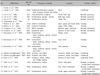
A literature search revealed 19 cases of fig leaves-induced phytophotodermatitis (Table 1)891011121314151617. Although cases following celery ingestion have been reported, most cases of phytophotodermatitis result from external contact18. Cases of fig leaves-induced phytophotodermatitis revealed that the plant was used for treatment, tanning, and gardening. In the cases reported here, patients used a fig leaves decoction for their underlying dermatologic conditions. Careful history taking is important to identify this unique phototoxic reaction, because it may be misdiagnosed as irritant contact dermatitis, allergic contact dermatitis, cellulitis, or tinea.
Although phytophotodermatitis from fig leaves has been reported, it is necessary to be aware of the folk remedies by unknown etiology.
In Korea, an old remedy for onychomycosis involves soaking feet in a vinegar solution. The use of fig leaves decoction for the treatment of underlying dermatologic diseases has also been shown. As for the cases reported here, several alternative medicines have no scientific basis and could lead to significant sequelae. As dermatologists, we should remind patients that phytophotodermatitis caused by fig leaves contact has no scientific basis, but present severe adverse events.
 XML Download