

 PDF
PDF ePub
ePub Citation
 Print
Print


INTRODUCTION
Amyloidosis can be subdivided into cutaneous amyloidosis and systemic amyloidosis with cutaneous involvement12. Amyloid precipitates are seen in the extracellular space of the dermis in both groups. Amyloid accumulates in the papillary dermis in patients with cutaneous amyloidosis, whereas it accumulates in subpapillary layers, dermal appendages, and blood vessels in patients with systemic amyloidosis and cutaneous involvement34. Secondary cutaneous amyloidosis refers to clinically unapparent amyloid deposits within the skin in association with preexisting skin conditions or skin tumors, such as basal cell carcinoma, porokeratosis, seborrheic keratosis, solar elastosis, Bowen's disease, and mycosis fungoides56789. It has also been reported following psoralen and ultraviolet A radiation (PUVA) therapy1011. We report an interesting and rare case of secondary cutaneous amyloidosis that occurred in patient with mycosis fungoides who was treated with PUVA and narrow band ultraviolet B (UVB) therapy.
CASE REPORT
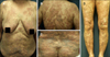
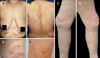
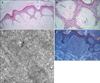
A 70-year-old woman presented with a 6-month history of asymptomatic multiple yellowish plaques on both lower extremities, including thighs and legs. She had been diagnosed with mycosis fungoides 7 years ago and had been treated with PUVA therapy, narrow-band UVB therapy, and acitretin medication for 5 years (Fig. 1). She had been treated with PUVA therapy firstly for 25 months and then treated with UVB therapy for 21 months. The phototherapy was done once a week or twice a week. The total number of exposure of PUVA was 72 and the cumulative PUVA radiation dose was 216.9 J/cm2. The total number of exposure of UVB was 98 and the cumulative UVB radiation dose was 110.35 J/cm2. Finally, she reached complete remission of mycosis fungoides (Fig. 2A, B). However, new yellowish lesions started to appear 1 year after discontinuing the phototherapy. A physical examination revealed multiple yellowish plaques on both lower extremities (Fig. 2C, D). The plaques were well circumscribed and slightly elevated (Fig. 2 E, F). Laboratory tests, including a complete blood cell count, differential leukocyte count, erythrocyte sedimentation rate, and blood chemistry studies were all normal. A biopsy specimen showed multiple nodular deposits of eosinophilic amorphous material in papillary dermis and upper reticular dermis (Fig. 3A, B). The deposits represented apple green birefringence on Congo red stain viewed under polarized light microscopy (Fig. 3C). The acellular small nodules in the upper dermis consisted of randomly oriented, non-branching, non-anastomosing 6.67~12.7 nm thick amyloid fibrils on electron microscopy (Fig. 3D). Therefore, we confirmed the diagnosis of secondary cutaneous amyloidosis. We reviewed past biopsy slides to determine when the amyloid had formed. Dense atypical lymphocyte infiltration in the dermis and epidermotrophism without evidence of amyloidosis were observed at the mycosis fungoides diagnosis 7 years ago. We found small multifocal deposits of faintly eosinophilic amorphous material confined to the papillary dermis but no evidence of mycosis fungoides 2 years ago during complete remission of the mycosis fungoides.
DISCUSSION
Localized amyloidosis refers to single organ-limited amyloid deposition. Localized cutaneous amyloidosis can occur as a primary phenomenon as in lichen or macular amyloidosis, or as a secondary phenomenon associated with another cutaneous pathology. Although amyloid deposits in different clinicopathological types of amyloidosis have histologically and morphologically identical properties, it has become apparent that there are several mechanisms of amyloid accumulation112.
In secondary amyloidosis associated with chronic inflammatory conditions, such as hidradenitis suppurativa, the amyloid protein precursors are acute phase reactants. However the deposits in localized amyloidosis are likely proteins synthesized within the affected tissue. Secondary localized cutaneous amyloidosis is usually associated with tumors of epidermal origin, and the amyloid is thought to be derived from keratinocytes11213. Although the precursor proteins of cutaneous amyloidosis have not been fully characterized, they are predominantly derived from keratinocytes in cutaneous amyloidosis. Converting a precursor to amyloid requires a conformational switch from α-pleated sheet arrangement to a β-pleated sheet structure. It is postulated that keratin tonofilaments undergo “filamentous degeneration” and keratinocytes “drop off” into the dermis forming amyloid13. A commonly accepted pathogenic theory of amyloidosis is that apoptotic basal keratinocytes (colloid bodies) release cytokeratins, which are covered with autoantibodies, phagocytized by macrophages, and enzymatically degraded into amyloid K (keratin-associated amyloid), which is a key feature of organ- limited cutaneous amyloidosis1314.
In our case, the clinical and histological features of cutaneous amyloidosis were observed without any clinical or histological features of mycosis fungoides. Secondary amyloid deposits can originate in association with mycosis fungoides. Several cases of secondary cutaneous amyloidosis associated with mycosis fungoides have been reported without any history of phototherapy8915. Izumi et al.15 reported that CD8+ T cells in poikilodermatous mycosis fungoides might be attributable to the formation of amyloid material from attacks on epidermal keratinocytes. The other theory associated with mycosis fungoides is that amyloid forms through prolonged scratching and rubbing, such as in friction amyloidosis. This theory is also supported by the fact that this patient complained of severe pruritus at the diagnosis of mycosis fungoides. A possible alternative mechanism of this case is amyloid formation through a cytotoxic effect of PUVA therapy. Secondary cutaneous amyloidosis was detected in five of 61 patients with mycosis fungoides simultaneously after PUVA by Zemheri et al.14. The mechanism of PUVA is phototoxic reactions resulting from direct cellular damage caused by an inflammatory, non-immunological mechanism. PUVA primarily targets DNA, and other important targets of psoralens are specific receptors, such as epidermal growth factor receptor. More recently, PUVA therapy can induce programmed cell death (apoptosis) in skin infiltrating T-helper lymphocytes and keratinocytes, so it cause interface changes101114. Lastly, we can also consider that amyloidosis might develop de novo without relation with mycosis fungoides or PUVA therapy. However, clinical features of amyloidosis in this patient did not correspond to primary amyloidosis such as lichen or macular amyloidosis. In addition, amyloidosis occurred at multiple sites along the both lower extremities, including thighs, knees, and lower legs. Accordingly, we think that amyloidosis in this patient could develop secondary to mycosis fungoides or PUVA therapy rather than de novo. We report an interesting and rare case of secondary cutaneous amyloidosis that developed during PUVA therapy and progressed for more than 1 year even though PUVA therapy was discontinued and mycosis fungoides was in complete remission.
 XML Download