

 PDF
PDF ePub
ePub Citation
 Print
Print


Abstract
Ankle injury is one of the most common injuries, and osteochondral lesions of the talus occur in up to 70% of acute ankle sprains or fractures. The number of sports injuries have increased due to the increase in leisure activities, and the development of diagnostic techniques to evaluate the cartilage status leads to a higher prevalence of osteochondral lesions of the talus. Although osteochondral lesions of the talus with no symptoms can be treated conservatively, adult patients are usually treated by surgery because they are more likely to fail after non-surgical management. Recovery to normal cartilage is important, but there has been no surgical treatment established for effective cartilage regeneration. Bone marrow stimulation, such as arthroscopic microfracture, is a commonly used surgical procedure and an effective treatment for lesions that are small or failed after non-operative treatment. In addition, there are treatments, such as osteochondral autograft transplantation, osteochondral allograft transplantation and autologous chondrocyte implantation. The selection of the methods depends on the size and location of the lesion, the presence of subchondral cysts, and the results of previous surgery. Many surgical procedures have shown good results in short and mid-term follow-up studies but the results of long-term followup have been unclear. Various treatment methods, such as hyaluronan, platelet-rich plasma, mesenchymal stem cells, and bone marrow aspirate concentrate, have been available recently due to the development of various biological agents.
REFERENCES
1. Canale ST, Belding RH. Osteochondral lesions of the talus. J Bone Joint Surg Am. 1980; 62:97–102.

2. Ulrich-Vinther M, Maloney MD, Schwarz EM, Rosier R, O'Keefe RJ. Articular cartilage biology. J Am Acad Orthop Surg. 2003; 11:421–30.
3. Mulfinger GL, Trueta J. The blood supply of the talus. J Bone Joint Surg Br. 1970; 52:160–7.
4. Verhagen RA, Struijs PA, Bossuyt PM, van Dijk CN. Systematic review of treatment strategies for osteochondral defects of the talar dome. Foot Ankle Clin. 2003; 8:233–42. viii-ix.
5. Mankin HJ. The response of articular cartilage to mechanical injury. J Bone Joint Surg Am. 1982; 64:460–6.
6. Berndt AL, Harty M. Transchondral fractures (osteochondritis dissecans) of the talus. J Bone Joint Surg Am. 1959; 41:988–1020.
7. Schachter AK, Chen AL, Reddy PD, Tejwani NC. Osteochondral lesions of the talus. J Am Acad Orthop Surg. 2005; 13:152–8.
8. Dekker TJ, Dekker PK, Tainter DM, Easley ME, Adams SB. Treatment of osteochondral lesions of the talus: a critical analysis review. JBJS Rev [Internet]. 2017. [cited 2017 Mar 28];5. doi: 10.2106/JBJS.RVW.16.00065.
9. McCoy AM, Toth F, Dolvik NI. . Articular osteochondrosis: a comparison of naturally-occurring human and animal disease. Osteoarthritis Cartilage. 2013; 21:1638–47.
10. van Dijk CN, Reilingh ML, Zengerink M, van Bergen CJ. Osteochondral defects in the ankle: why painful? Knee Surg Sports Traumatol Arthrosc. 2010; 18:570–80.
11. Tol JL, Struijs PA, Bossuyt PM, Verhagen RA, van Dijk CN. Treatment strategies in osteochondral defects of the talar dome: a systematic review. Foot Ankle Int. 2000; 21:119–26.
12. Alexander AH, Lichtman DM. Surgical treatment of transchondral talar-dome fractures (osteochondritis dissecans). Long-term follow-up. J Bone Joint Surg Am. 1980; 62:646–52.
13. Leontaritis N, Hinojosa L, Panchbhavi VK. Arthroscopically detected intra-articular lesions associated with acute ankle fractures. J Bone Joint Surg Am. 2009; 91:333–9.
14. Saxena A, Eakin C. Articular talar injuries in athletes: results of microfracture and autogenous bone graft. Am J Sports Med. 2007; 35:1680–7.
15. O'Loughlin PF, Heyworth BE, Kennedy JG. Current concepts in the diagnosis and treatment of osteochondral lesions of the ankle. Am J Sports Med. 2010; 38:392–404.
16. Hembree WC, Wittstein JR, Vinson EN. . Magnetic resonance imaging features of osteochondral lesions of the talus. Foot Ankle Int. 2012; 33:591–7.
17. Chew KT, Tay E, Wong YS. Osteochondral lesions of the talus. Ann Acad Med Singapore. 2008; 37:63–8.
18. Verhagen RA, Maas M, Dijkgraaf MG, Tol JL, Krips R, van Dijk CN. Prospective study on diagnostic strategies in osteochondral lesions of the talus. Is MRI superior to helical CT? J Bone Joint Surg Br. 2005; 87:41–6.
19. Canale ST, Kelly FB Jr. Fractures of the neck of the talus. Long-term evaluation of seventy-one cases. J Bone Joint Surg Am. 1978; 60:143–56.
20. Loomer R, Fisher C, Lloyd-Smith R, Sisler J, Cooney T. Osteochondral lesions of the talus. Am J Sports Med. 1993; 21:13–9.
21. Adams SB, Parekh SG, de Solminihac DHZ, Krynetskiy EE, Schon LC, Easley ME. Cartilage repair, replacement, and regenerative strategies for osteochondral lesions of the talus. 2014. London: Springer;p. 269–93.
22. Anderson IF, Crichton KJ, Grattan-Smith T, Cooper RA, Brazier D. Osteochondral fractures of the dome of the talus. J Bone Joint Surg Am. 1989; 71:1143–52.
23. Ferkel RD, Flannigan BD, Elkins BS. Magnetic resonance imaging of the foot and ankle: correlation of normal anatomy with pathologic conditions. Foot Ankle. 1991; 11:289–305.
24. Mintz DN, Tashjian GS, Connell DA, Deland JT, O'Malley M, Potter HG. Osteochondral lesions of the talus: a new magnetic resonance grading system with arthroscopic correlation. Arthroscopy. 2003; 19:353–9.
25. Pritsch M, Horoshovski H, Farine I. Arthroscopic treatment of osteochondral lesions of the talus. J Bone Joint Surg Am. 1986; 68:862–5.
26. Elias I, Jung JW, Raikin SM, Schweitzer MW, Carrino JA, Morrison WB. Osteochondral lesions of the talus: change in MRI findings over time in talar lesions without operative intervention and implications for staging systems. Foot Ankle Int. 2006; 27:157–66.
27. Easley ME. Scranton PE Jr. Osteochondral autologous transfer system. Foot Ankle Clin. 2003; 8:275–90.
28. Edmonds EW, Shea KG. Osteochondritis dissecans: editorial comment. Clin Orthop Relat Res. 2013; 471:1105–6.
29. Bauer M, Jonsson K, Lindén B. Osteochondritis dissecans of the ankle. A 20-year follow-up study. J Bone Joint Surg Br. 1987; 69:93–6.
30. Hannon CP, Smyth NA, Murawski CD. . Osteochondral lesions of the talus: aspects of current management. Bone Joint J. 2014; 96:164–71.
31. Klammer G, Maquieira GJ, Spahn S, Vigfusson V, Zanetti M, Espinosa N. Natural history of nonoperatively treated osteochondral lesions of the talus. Foot Ankle Int. 2015; 36:24–31.
32. Zengerink M, Struijs PA, Tol JL, van Dijk CN. Treatment of osteochondral lesions of the talus: a systematic review. Knee Surg Sports Traumatol Arthrosc. 2010; 18:238–46.
33. Shearer C, Loomer R, Clement D. Nonoperatively managed stage 5 osteochondral talar lesions. Foot Ankle Int. 2002; 23:651–4.
34. Mei-Dan O, Carmont MR, Laver L, Mann G, Maffulli N, Nyska M. Platelet-rich plasma or hyaluronate in the management of osteochondral lesions of the talus. Am J Sports Med. 2012; 40:534–41.
35. Giannini S, Buda R, Faldini C. . Surgical treatment of osteochondral lesions of the talus in young active patients. J Bone Joint Surg Am. 2005; (87 Suppl):2:28–41.
36. Gobbi A, Francisco RA, Lubowitz JH, Allegra F, Canata G. Osteochondral lesions of the talus: randomized controlled trial comparing chondroplasty, microfracture, and osteochondral autograft transplantation. Arthroscopy. 2006; 22:1085–92.
37. Loveday D, Clifton R, Robinson A. Interventions for treating osteochondral defects of the talus in adults. Cochrane Database Syst Rev. 2010; 8:CD00–8104.
38. Anderson AF, Pagnani MJ. Osteochondritis dissecans of the femoral condyles. Long-term results of excision of the fragment. Am J Sports Med. 1997; 25:830–4.
39. O'Driscoll SW. The healing and regeneration of articular cartilage. J Bone Joint Surg Am. 1998; 80:1795–812.
40. Larsen MW, Pietrzak WS, DeLee JC. Fixation of osteochondritis dissecans lesions using poly(l-lactic acid)/ poly(glycolic acid) copolymer bioabsorbable screws. Am J Sports Med. 2005; 33:68–76.
41. Taranow WS, Bisignani GA, Towers JD, Conti SF. Retrograde drilling of osteochondral lesions of the medial talar dome. Foot Ankle Int. 1999; 20:474–80.
42. Kono M, Takao M, Naito K, Uchio Y, Ochi M. Retrograde drilling for osteochondral lesions of the talar dome. Am J Sports Med. 2006; 34:1450–6.
43. Ferkel RD, Zanotti RM, Komenda GA. . Arthroscopic treatment of chronic osteochondral lesions of the talus: longterm results. Am J Sports Med. 2008; 36:1750–62.
44. Anders S, Lechler P, Rackl W, Grifka J, Schaumburger J. Fluoroscopy-guided retrograde core drilling and cancellous bone grafting in osteochondral defects of the talus. Int Orthop. 2012; 36:1635–40.
45. Nehrer S, Spector M, Minas T. Histologic analysis of tissue after failed cartilage repair procedures. Clin Orthop Relat Res. 1999; 365:149–62.
46. Becher C, Thermann H. Results of microfracture in the treatment of articular cartilage defects of the talus. Foot Ankle Int. 2005; 26:583–9.
47. Polat G, Erşen A, Erdil ME, Kızılkurt T; Kılıçoğlu Ö, Aşık M. Long-term results of microfracture in the treatment of talus osteochondral lesions. Knee Surg Sports Traumatol Arthrosc. 2016; 24:1299–303.
48. Chuckpaiwong B, Berkson EM, Theodore GH. Microfracture for osteochondral lesions of the ankle: outcome analysis and outcome predictors of 105 cases. Arthroscopy. 2008; 24:106–12.
49. Choi WJ, Park KK, Kim BS, Lee JW. . Osteochondral lesion of the talus: is there a critical defect size for poor outcome? Am J Sports Med. 2009; 37:1974–80.
50. Ogilvie-Harris DJ, Sarrosa EA. Arthroscopic treatment of post-traumatic cysts of the talus. Arthroscopy. 2000; 16:197–201.
51. Savva N, Jabur M, Davies M, Saxby T. Osteochondral lesions of the talus: results of repeat arthroscopic debridement. Foot Ankle Int. 2007; 28:669–73.
52. Lee KB, Park HW, Cho HJ, Seon JK. Comparison of ar-throscopic microfracture for osteochondral lesions of the talus with and without subchondral cyst. Am J Sports Med. 2015; 43:1951–6.
53. Hepple S, Winson IG, Glew D. Osteochondral lesions of the talus: a revised classification. Foot Ankle Int. 1999; 20:789–93.
54. Hu Y, Guo Q, Jiao C. . Treatment of large cystic medial osteochondral lesions of the talus with autologous osteoperiosteal cylinder grafts. Arthroscopy. 2013; 29:1372–9.
55. Radin EL, Rose RM. Role of subchondral bone in the initiation and progression of cartilage damage. Clin Orthop Relat Res. 1986; 213:34–40.
56. Shimozono Y, Coale M, Yasui Y, O'Halloran A, Deyer TW, Kennedy JG. Subchondral bone degradation after microfracture for osteochondral lesions of the talus: an MRI analysis. Am J Sports Med. 2018; 46:642–8.
57. Reilingh ML, van Bergen CJ, Blankevoort L, et al. Computed tomography analysis of osteochondral defects of the talus after arthroscopic debridement and microfracture. Knee Surg Sports Traumatol Arthrosc. 2016; 24:1286–92.
58. Murawski CD, Duke GL, Deyer TW, Kennedy JG. Bone Marrow Aspirate Concentrate (BMAC) as a biological adjunct to the surgical treatment of osteochondral lesions of the talus. Tech Foot Ankle Surg. 2011; 10:18–27.
59. Fortier LA, Potter HG, Rickey EJ. . Concentrated bone marrow aspirate improves full-thickness cartilage repair compared with microfracture in the equine model. J Bone Joint Surg Am. 2010; 92:1927–37.
60. Hannon CP, Ross KA, Murawski CD. . Arthroscopic bone marrow stimulation and concentrated bone marrow aspirate for osteochondral lesions of the talus: a case-control study of functional and magnetic resonance observation of cartilage repair tissue outcomes. Arthroscopy. 2016; 32:339–47.
61. Ogilvie-Harris DJ, Sarrosa EA. Arthroscopic treatment after previous failed open surgery for osteochondritis dissecans of the talus. Arthroscopy. 1999; 15:809–12.
62. Yoon HS, Park YJ, Lee M, Choi WJ, Lee JW. Osteochondral autologous transplantation is superior to repeat arthroscopy for the treatment of osteochondral lesions of the talus after failed primary arthroscopic treatment. Am J Sports Med. 2014; 42:1896–903.
63. McGahan PJ, Pinney SJ. Current concept review: osteochondral lesions of the talus. Foot Ankle Int. 2010; 31:90–101.
64. Sasaki K, Ishibashi Y, Sato H, Toh S. Arthroscopically assisted osteochondral autogenous transplantation for osteochondral lesion of the talus using a transmalleolar approach. Arthros-copy. 2003; 19:922–7.
65. Tochigi Y, Amendola A, Muir D, Saltzman C. Surgical approach for centrolateral talar osteochondral lesions with an anterolateral osteotomy. Foot Ankle Int. 2002; 23:1038–9.
66. Imhoff AB, Paul J, Ottinger B. . Osteochondral transplantation of the talus: long-term clinical and magnetic resonance imaging evaluation. Am J Sports Med. 2011; 39:1487–93.
67. Valderrabano V, Leumann A, Rasch H, Egelhof T, Hinter-mann B, Pagenstert G. Knee-to-ankle mosaicplasty for the treatment of osteochondral lesions of the ankle joint. Am J Sports Med. 2009; 37:105S–11S.
68. Kim YS, Park EH, Kim YC, Koh YG, Lee JW. Factors associated with the clinical outcomes of the osteochondral autograft transfer system in osteochondral lesions of the talus: second-look arthroscopic evaluation. Am J Sports Med. 2012; 40:2709–19.
69. Woelfle JV, Reichel H, Nelitz M. Indications and limitations of osteochondral autologous transplantation in osteochondritis dissecans of the talus. Knee Surg Sports Traumatol Ar-throsc. 2013; 21:1925–30.
70. Emre TY, Ege T, Cift HT, Demircioğlu DT, Seyhan B, Uzun M. Open mosaicplasty in osteochondral lesions of the talus: a prospective study. J Foot Ankle Surg. 2012; 51:556–60.
71. Scranton PE Jr, Frey CC, Feder KS. Outcome of osteochondral autograft transplantation for type-V cystic osteochondral lesions of the talus. J Bone Joint Surg Br. 2006; 88:614–9.
72. Murawski CD, Kennedy JG. Operative treatment of osteochondral lesions of the talus. J Bone Joint Surg Am. 2013; 95:1045–54.
73. Williams SK, Amiel D, Ball ST. . Prolonged storage effects on the articular cartilage of fresh human osteochondral allografts. J Bone Joint Surg Am. 2003; 85:2111–20.
74. Gross CE, Adams SB, Easley ME, Nunley JA 2nd. Role of fresh osteochondral allografts for large talar osteochondral lesions. J Am Acad Orthop Surg. 2016; 24:e9–17.
75. El-Rashidy H, Villacis D, Omar I, Kelikian AS. Fresh osteochondral allograft for the treatment of cartilage defects of the talus: a retrospective review. J Bone Joint Surg Am. 2011; 93:1634–40.
76. Adams SB Jr, Viens NA, Easley ME, Stinnett SS, Nunley JA 2nd. Midterm results of osteochondral lesions of the talar shoulder treated with fresh osteochondral allograft transplantation. J Bone Joint Surg Am. 2011; 93:648–54.
77. Mandelbaum BR, Gerhardt MB, Peterson L. Autologous chondrocyte implantation of the talus. Arthroscopy. 2003; 19(Suppl 1):129–37.
78. Giannini S, Buda R, Vannini F, Di Caprio F, Grigolo B. Ar-throscopic autologous chondrocyte implantation in osteochondral lesions of the talus: surgical technique and results. Am J Sports Med. 2008; 36:873–80.
79. Niemeyer P, Salzmann G, Schmal H, Mayr H, Südkamp NP. Autologous chondrocyte implantation for the treatment of chondral and osteochondral defects of the talus: a meta-analysis of available evidence. Knee Surg Sports Traumatol Ar-throsc. 2012; 20:1696–703.
80. Baums MH, Heidrich G, Schultz W, Steckel H, Kahl E, Klinger HM. Autologous chondrocyte transplantation for treating cartilage defects of the talus. J Bone Joint Surg Am. 2006; 88:303–8.
81. Buda R, Vannini F, Castagnini F, Cavallo M, Ruffilli A, Ram-poni L. . Regenerative treatment in osteochondral lesions of the talus: autologous chondrocyte implantation versus one-step bone marrow derived cells transplantation. Int Or-thop. 2015; 39:893–900.
82. Nam EK, Ferkel RD, Applegate GR. Autologous chondrocyte implantation of the ankle: a 2- to 5-year follow-up. Am J Sports Med. 2009; 37:274–84.
83. Battaglia M, Vannini F, Buda R. . Arthroscopic autologous chondrocyte implantation in osteochondral lesions of the talus: mid-term T2-mapping MRI evaluation. Knee Surg Sports Traumatol Arthrosc. 2011; 19:1376–84.
84. Kwak SK, Kern BS, Ferkel RD, Chan KW, Kasraeian S, Applegate GR. Autologous chondrocyte implantation of the ankle: 2- to 10-year results. Am J Sports Med. 2014; 42:2156–64.
85. Brittberg M, Peterson L, Sjögren-Jansson E, Tallheden T, Lindahl A. Articular cartilage engineering with autologous chondrocyte transplantation. A review of recent developments. J Bone Joint Surg Am. 2003; 85(Suppl 3):109–15.
86. Giza E, Sullivan M, Ocel D. . Matrix-induced autologous chondrocyte implantation of talus articular defects. Foot Ankle Int. 2010; 31:747–53.
87. Valderrabano V, Miska M, Leumann A, Wiewiorski M. Reconstruction of osteochondral lesions of the talus with autologous spongiosa grafts and autologous matrix-induced chondrogenesis. Am J Sports Med. 2013; 41:519–27.
88. Kreulen C, Giza E, Walton J, Sullivan M. Seven-year follow-up of matrix-induced autologous implantation in talus articular defects. Foot Ankle Spec. 2018; 11:133–7.
Figure 1
Simple radiograph and ar-throscopic view of the osteochondral lesion of the talus. (A) Simple radiograph shows radiolucent lesion at medial talus. (B) Arthroscopic view.
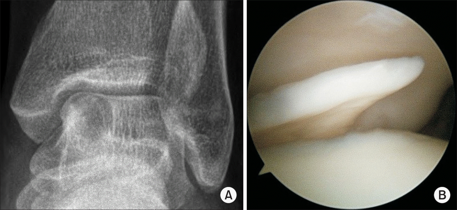
Figure 2
Arthroscopic microfracture. (A) Arthroscopic view of the osteochondral lesion of the talus. (B) Debridement and curettage was performed at the osteochondral lesion. (C) Microfracture was performed. (D) Bleeding after microfracture.
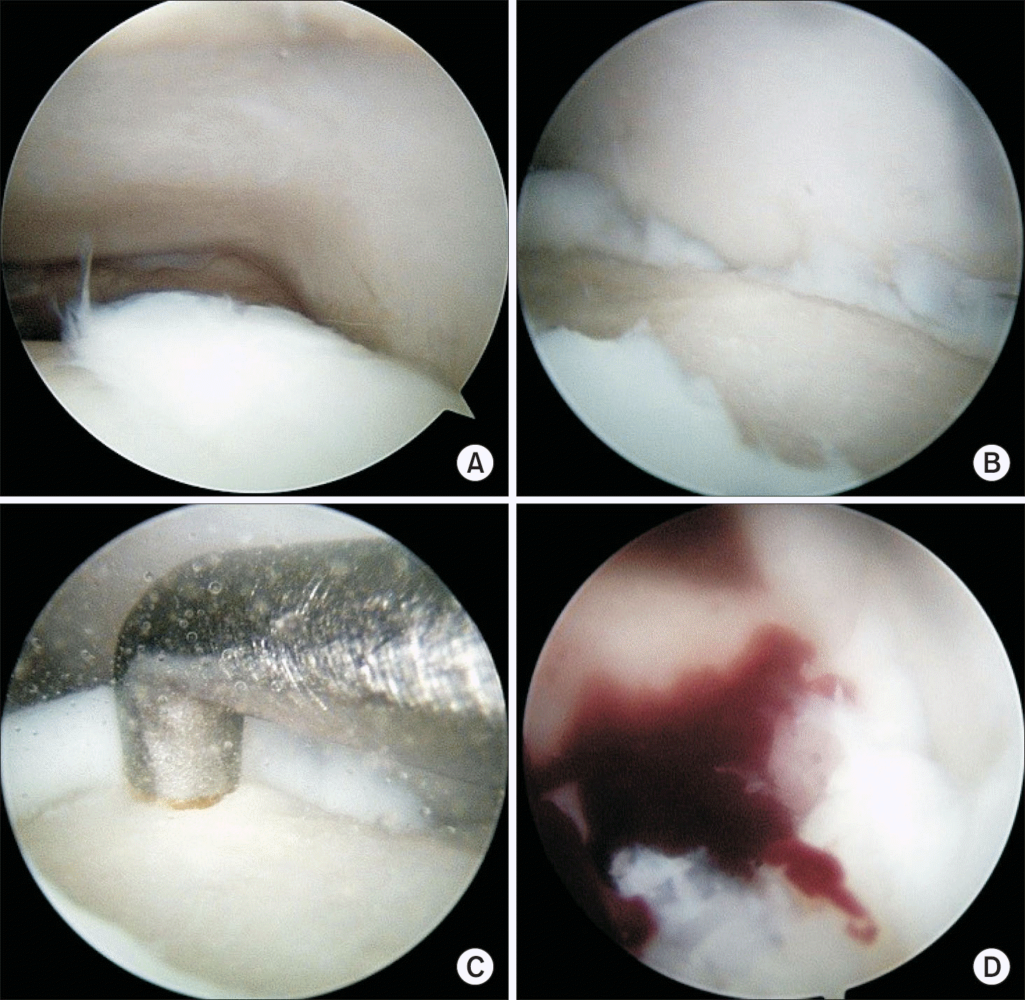
Figure 3
Osteochondral autologous transplantation. (A) Preoperative magnetic resonance imaging. (B) Preparation of the talus through medial malleolar osteotomy. (C) Autologous osteochondral harvest from lateral femomal condyle. (D) Osteochondral autologous transplantation was performed. (E) Secondary arthroscopic view after 2 years.
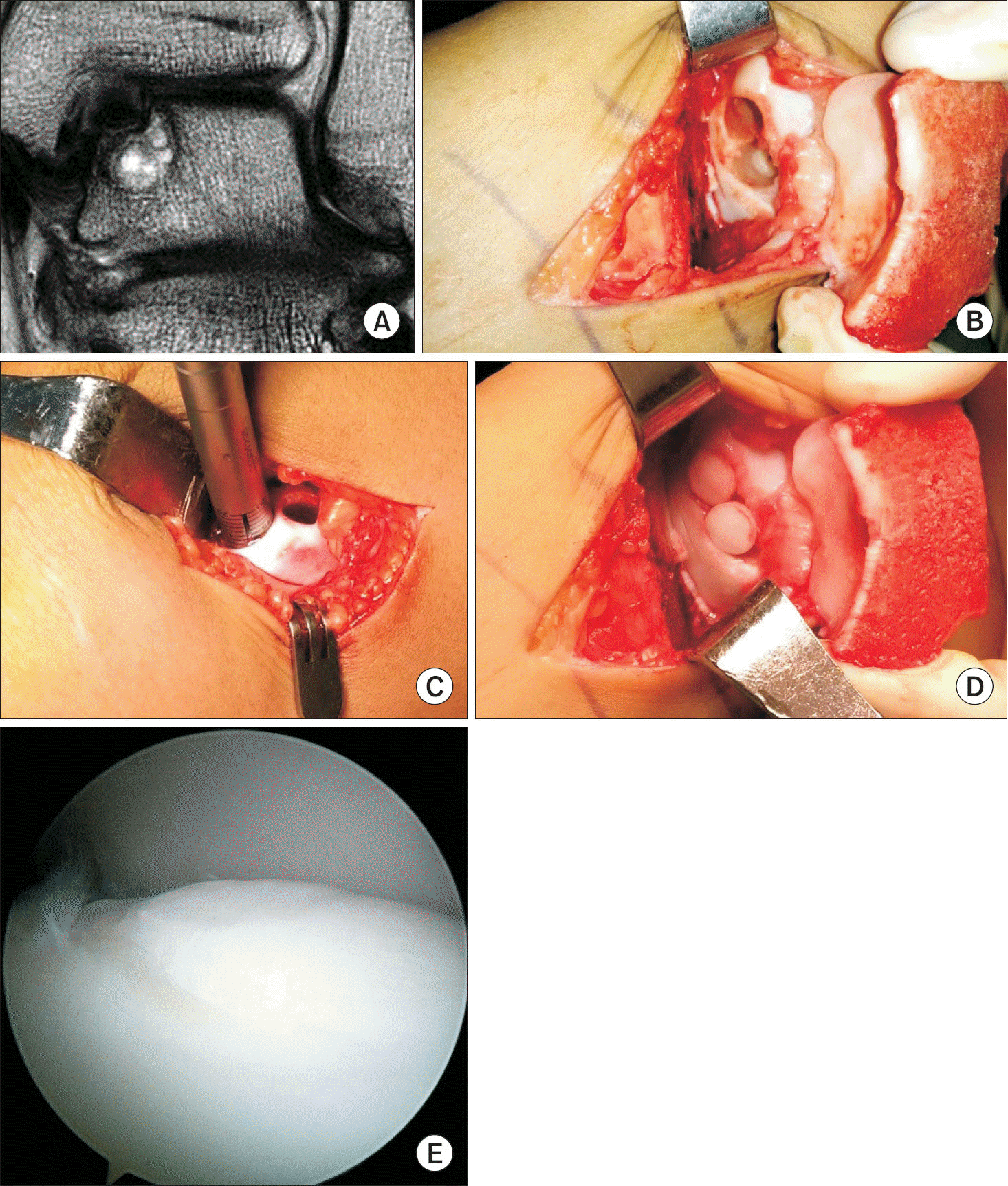
Table 1
Various Classification of the OLT
 XML Download