

 PDF
PDF ePub
ePub Citation
 Print
Print


Abstract
Purpose
Clear cell chondrosarcoma may have a benign appearance even on a magnetic resonance imaging (MRI). Hence, it can be confused with benign bone tumors, such as a giant cell tumor or chondroblastoma. The aim of our study was to document the doctor-associated diagnostic errors in patients with clear cell chondrosarcoma and oncologic outcomes of these lesions, which were misdiagnosed as benign bone tumors.
Materials and Methods
We identified 10 patients who were diagnosed with and treated for clear cell chondrosarcoma between January 1996 and December 2014. One patient was excluded due to insufficient clinical data. We then reviewed their data regarding age, gender, symptom onset, tumor location, initial imaging diagnosis, and associated previous treatment. We examined the errors of surgeons and pathologists with respect to patient and tumor characteristics. We also analyzed treatment delay, time to local recurrence, metastasis, follow-up duration, and the oncologic outcome.
Results
The initial presumptive diagnosis based on MRIs for all 9 patients was benign bone tumor. Among 8 patients who underwent inappropriate procedure, half of them were diagnosed as clear cell chondrosarcoma immediately after the curettage. As for the remaining 4 patients, the surgeon did not send any tissue samples to a pathologist for a definite diagnosis in three patients and a pathologist made an incorrect diagnosis in one patient. We performed an appropriate surgery on all patients with a wide surgical margin. The average treatment delay was 27 months (range, 0–127 months), and the average follow-up duration was 65 months (range, 13–164 months). One patient had local recurrence after 12 months. Metastatic disease developed in 2 patients with a median time to definitive treatment of 24 months (12–37 months). Ten-year overall survival of patients with clear cell chondrosarcoma was 78%, and two patients died due to disease progression.
Conclusion
Misdiagnosis of clear cell chondrosacroma as a benign bone tumor is not uncommon, even for experienced orthopaedic oncologists, resulting in definite curative surgery without biopsy. An inappropriate primary treatment may increase the risk of local recurrence and metastasis. Therefore, a proper subsequent surgery is mandatory for patients with clear cell chondrosarcoma who received inadvertent curettage.
Figures and Tables
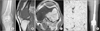 | Figure 1A 58-year-old man with left knee pain that developed 4 days ago after a slip down (case No. 5). (A) Initial plain radiograph shows a well marginated benign-looking bone lesion at the distal femur epiphysis. (B, C) Sagittal and axial T1 weighted images show intraosseous bone lesion with subtle cortical involvement. Initial presumptive diagnosis based on images was giant cell tumor. (D) Incisional biopsy revealed a typical feature of clear cell chondrosarcoma, which showed a clear cell, with a large, extensive cytoplasm with distinct cell boundary (H&E, ×100). (E) Wide excision and reconstruction using tumor prosthesis were performed.
|
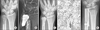 | Figure 2A 32-year-old man with right wrist pain (case No. 3). (A) Initial plain radiograph shows well-marginated benign looking bone lesion at the distal ulna. (B) Coronal enhanced T1 weighted image shows intraosseous bone lesion with subtle cortical involvement. Initial presumptive diagnosis based on image was a giant cell tumor. (C) Curettage and bone cementing were performed. (D) Specimen revealed that the diagnosis was clear cell chondrosarcoma (H&E, ×400). (E) Wide excision and reconstruction using overlapping allograft were performed. To enhance the forearm function of the patient, we used an allograft reconstruction instead of resection only.
|
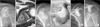 | Figure 3An 8-year-old girl with left shoulder pain (case No. 7). (A) Initial plain radiograph shows bone lesion at the left scapula. (B) Axial T2 weighted image shows a bone lesion with subtle cortical involvement. Initial presumptive diagnosis based on images was chondroblastoma. (C) Curettage and bone graft was performed at referral hospital. Diagnosis was clear cell chondrosarcoma. (D) Wide excision and reconstruction using pasteurized bone with bone cement was performed at our hospital. (E) Postoperative 3-month plain radiograph shows the reconstructed scapula using pasteurized autograft. (F) Postoperative 6-year plain radiograph shows total resorption of the pasteurized bone with deformed shoulder joint. However, she has continuously been disease free with no disease relapse.
|
Notes
References
1. Unni KK, Dahlin DC, Beabout JW, Sim FH. Chondrosarcoma: clear-cell variant. A report of sixteen cases. J Bone Joint Surg Am. 1976; 58:676–683.
2. Bjornsson J, Unni KK, Dahlin DC, Beabout JW, Sim FH. Clear cell chondrosarcoma of bone. Observations in 47 cases. Am J Surg Pathol. 1984; 8:223–230.
3. Yamaguchi H, Isu K, Ubayama Y, et al. Clear cell chondrosarcoma. A report of two cases and review of literature. Acta Pathol Jpn. 1986; 36:1577–1585.

4. Donati D, Yin JQ, Colangeli M, et al. Clear cell chondrosarcoma of bone: long time follow-up of 18 cases. Arch Orthop Trauma Surg. 2008; 128:137–142.
5. Itälä A, Leerapun T, Inwards C, Collins M, Scully SP. An institutional review of clear cell chondrosarcoma. Clin Orthop Relat Res. 2005; 440:209–212.
6. Collins MS, Koyama T, Swee RG, Inwards CY. Clear cell chondrosarcoma: radiographic, computed tomographic, and magnetic resonance findings in 34 patients with pathologic correlation. Skeletal Radiol. 2003; 32:687–694.
7. Jeong HJ, Ko SK, Park MS, Chang HK, Huh MH. Clear cell chondrosarcoma arising in hyoid bone. Korean J Pathol. 1997; 31:470–475.
8. Kang CM, Han CS, Jung GY, Jeong HY, Kim YJ. Clear cell chondrosarcoma of the tibia diaphysis: a case report. J Korean Bone Joint Tumor Soc. 2014; 20:89–93.
9. Lee K, Lee AH, Kim J, Kim HM, Lee KY. Clear cell chondrosarcoma of the scapula in a child: a case report. J Korean Bone Joint Tumor Soc. 2009; 15:155–159.
10. Lee SD, Ahn GH, Chi JG, Ham EK. Clear-cell chondrosarcoma--a case report. J Korean Med Sci. 1989; 4:155–158.
11. Lee YG, Lee M, Park GS, Ahn GH. Clear cell chondrosarcoma. J Korean Orthop Assoc. 1983; 18:419–421.
12. Mankin HJ, Mankin CJ, Simon MA. The hazards of the biopsy, revisited. Members of the musculoskeletal tumor society. J Bone Joint Surg Am. 1996; 78:656–663.
13. Markel DC, Neumann KU, Steinau HU. Appropriate techniques for musculoskeletal tumor biopsy. Orthop Rev. 1994; 23:176–180.
14. Bickels J, Jelinek JS, Shmookler BM, Neff RS, Malawer MM. Biopsy of musculoskeletal tumors. Current concepts. Clin Orthop Relat Res. 1999; 368:212–219.
15. Simon MA, Biermann JS. Biopsy of bone and soft-tissue lesions. J Bone Joint Surg Am. 1993; 75:616–621.
16. Ayoub KS, Grimer RJ, Carter SR, Mangham DC, Davies AM, Tillman RM. Clear cell chondrosarcoma of bone. Sarcoma. 1999; 3:115–119.
 XML Download