

 PDF
PDF ePub
ePub Citation
 Print
Print


Abstract
Hypereosinophilia, defined as an absolute eosinophil count of >1,500/μL, can be caused by a number of allergic, infectious, para-neoplastic and neoplastic disorders. In cases of hypereosinophilia with lymphoid proliferation, pathological confirmation is essential to exclude either myeloid or lymphoid malignancy. A 38-year-old woman with both cervical lymphadenopathies and peripheral blood eosinophilia visited our clinic. She had already performed core biopsy of lymph nodes and diagnosed as Kimura disease at a regional hospital. At the time of our clinic visit, there were no palpable cervical lymph nodes. The blood test showed hypereosinophilia with a high total IgE level. There was no evidence of tissue infiltration of eosinophils except for duodenitis with eosinophilic infiltration. Based on these findings, she was diagnosed as Kimura disease. She treated with high-dose systemic corticosteroid (1 mg/kg) and additional immunosuppressants sequentially used cyclophosphamide and cyclosporine. However, her eosinophilia waxed and waned, and a left inguinal mass was newly found. Excisional biopsy findings showed large atypical lymphoid cells with numerous eosinophilis, and immunohistochemistry showed CD3+, CD20-, CD30+ and anaplastic lymphoma kinase (ALK). The final diagnosis was ALK-negative anaplastic large cell lymphoma. We report a case of anaplastic large cell lymphoma with marked peripheral eosinophilia misdiagnosed as Kimura disease. In the case of hypereosinophilia with lymphadenopathy, it is necessary to differentiate hematologic diseases through immunochemical staining.
REFERENCES
1. Helbig G. Advances in the diagnosis and treatment of eosinophilia. Curr Opin Hematol. 2014; 21:3–7.

2. Gotlib J. World Health Organization-defined eosinophilic disorders: 2015 update on diagnosis, risk stratification, and management. Am J Hematol. 2015; 90:1077–89.
3. Sun QF, Xu DZ, Pan SH, Ding JG, Xue ZQ, Miao CS, et al. Kimura disease: review of the literature. Intern Med J. 2008; 38:668–72.
4. Chen H, Thompson LD, Aguilera NS, Abbondanzo SL. Kimura disease: a clinicopathologic study of 21 cases. Am J Surg Pathol. 2004; 28:505–13.
5. Valent P, Klion AD, Horny HP, Roufosse F, Gotlib J, Weller PF, et al. Con-temporary consensus proposal on criteria and classification of eosinophilic disorders and related syndromes. J Allergy Clin Immunol. 2012; 130:607–12.e9.
6. Hsieh FH. Hypereosinophilic syndrome. Ann Allergy Asthma Immunol. 2014; 112:484–8.
7. Park CS, Lee SP. Recent advances in the classification and management of hypereosinophilia. Allergy Asthma Respir Dis. 2015; 3:387–95.
8. Ogbogu PU, Bochner BS, Butterfield JH, Gleich GJ, Huss-Marp J, Kahn JE, et al. Hypereosinophilic syndrome: a multicenter, retrospective analysis of clinical characteristics and response to therapy. J Allergy Clin Immunol. 2009; 124:1319–25.e3.
9. Roche-Lestienne C, Lepers S, Soenen-Cornu V, Kahn JE, Laï JL, Hachulla E, et al. Molecular characterization of the idiopathic hypereosinophilic syndrome (HES) in 35 French patients with normal conventional cyto-genetics. Leukemia. 2005; 19:792–8.
10. Chihara D, Fanale MA. Management of anaplastic large cell lymphoma. Hematol Oncol Clin North Am. 2017; 31:209–22.
11. Mak V, Hamm J, Chhanabhai M, Shenkier T, Klasa R, Sehn LH, et al. Survival of patients with peripheral T-cell lymphoma after first relapse or progression: spectrum of disease and rare long-term survivors. J Clin Oncol. 2013; 31:1970–6.
12. Takimoto Y, Tanaka H, Tanabe O, Kuramoto A, Sasaki N, Nanba K. Anaplastic large-cell lymphoma (Ki-1 lymphoma) with expression of IL-5 mRNA and eosinophilic invasion. Acta Haematol. 1996; 96:245–8.
13. McCluggage WG, Walsh MY, Bharucha H. Anaplastic large cell malig-nant lymphoma with extensive eosinophilic or neutrophilic infiltration. Histopathology. 1998; 32:110–5.
14. McKelvie PA, Oon S, Romas E, Nandurkar H, Tam CS. A case of systemic anaplastic lymphoma kinase-negative anaplastic large cell lymphoma associated with hypereosinophilia, granulomatous myositis and vasculitis. Leuk Lymphoma. 2012; 53:2279–82.
15. Powers ML, Watson BW, Frater JL, Kreisel F, Hassan A. Peripheral eosinophilia camouflaging anaplastic large cell lymphoma. Int J Surg Pathol. 2011; 19:405–8.
16. Orofino N, Guidotti F, Cattaneo D, Sciumè M, Gianelli U, Cortelezzi A, et al. Marked eosinophilia as initial presentation of breast implant-associated anaplastic large cell lymphoma. Leuk Lymphoma. 2016; 57:2712–5.
17. Duval A, Bergoin E, Maynadié M, Bonnotte B, Girodon F. Images in hae-matology. Hypereosinophilia as a presenting feature of anaplastic large cell lymphoma. Br J Haematol. 2008; 140:363.
18. Choi W, Park YH, Paik KH, Chang YH, Lee SS, Ryoo BY, et al. Peripheral T-cell lymphoma-unspecified (PTCL-U) presenting with hypereosinophilic syndrome and pleural effusions. Korean J Intern Med. 2006; 21:57–61.
19. Kim CJ, Park SH, Chi JG. Idiopathic hypereosinophilic syndrome termi-nating as disseminated T-cell lymphoma. Cancer. 1991; 67:1064–9.
20. Koh YI. Clinical characteristics of Kimura's disease. Korean J Asthma Allergy Clin Immunol. 2010; 30:275–6.
Fig. 1.
Summary of peripheral eosinophil counts and the regimen of treatment. Clinical course of the patient from the initial visit to final diagnosis. LN, lymph node.
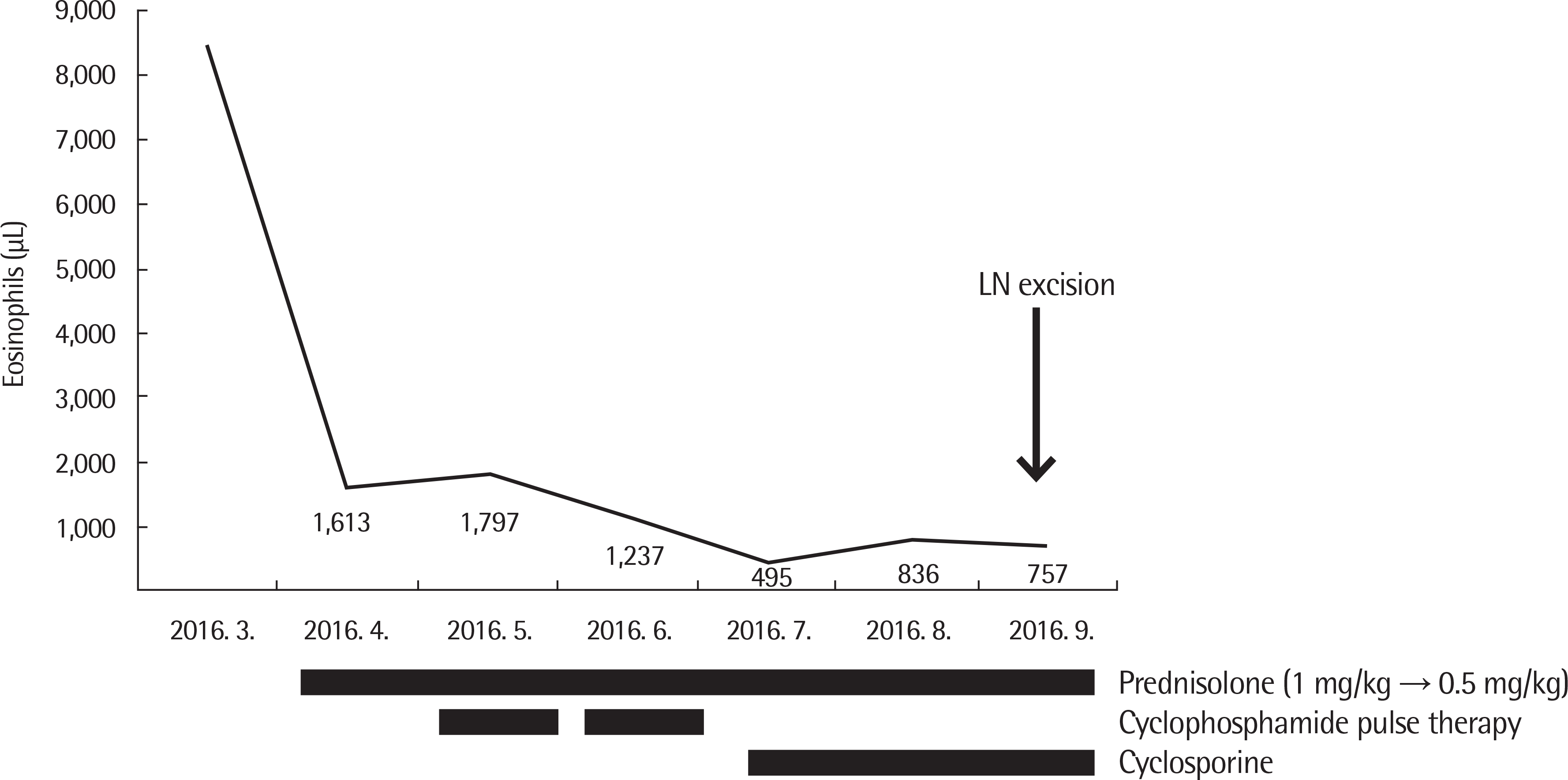
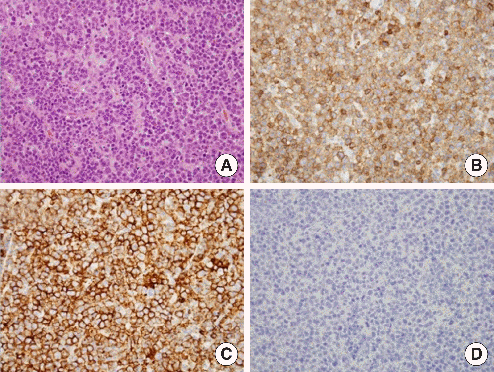
Fig. 2.
Pathologic findings of left inguinal lymph node. (A) Anaplastic large cell lymphoma showed marked hypercellularity with atypical cells with prominent nucleoli (H&E, ×200). (B) Positive staining of CD3 (×200). (C) Positive staining of CD30 (×200). (D) Negative staining of anaplastic lymphoma kinase (×200).
 XML Download