

 PDF
PDF ePub
ePub Citation
 Print
Print


INTRODUCTION
The clinical outcomes of extracorporeal membrane oxygenation (ECMO) show ongoing improvement, and ECMO treatment currently focuses on the control of complications.1 Nosocomial infections are among the major complications that increase patient mortality during ECMO.2 Unfortunately, the use of extracorporeal circulation itself may be a cause of systemic inflammatory response syndrome (SIRS). Therefore, it may be difficult to differentiate between infectious complications arising early after ECMO is initiated and the general inflammatory response to the use of extracorporeal circulation.
Various methods for the diagnosis of infection have been studied. Biomarkers such as white blood cells (WBCs) and C-reactive protein (CRP), in addition to blood cultures, have been used in diagnosis. Existing biomarkers in patients with suspected infection have limitations in sensitivity and specificity, and blood cultures are slow to identify organisms, with the possibility of false negatives. Procalcitonin (PCT) is the peptide precursor of the hormone calcitonin, and under normal circumstances is produced in the thyroid parafollicular cell (normal serum concentration <0.05 ng/mL). The PCT level has proven to be highly specific, and is a predictive marker for occurrence of bacterial infection and the decision to terminate antibiotic treatment in critically ill patients.34
However, in addition to infectious disease, PCT is also elevated in a variety of illnesses that cause inflammatory reactions such as complications associated with abdominal surgery, transplantation, trauma, and burns.345 Recently, its usefulness has been confirmed in non-infectious diseases such as ischemic stroke.6 An unusual increase in PCT, regardless of infection, has also been observed during ECMO support.78 We evaluated trends and predictive value of PCT levels measured during ECMO for prediction of clinical outcomes, such as infection and survival.
MATERIALS AND METHODS
1. Patient selection
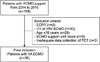
This retrospective study included consecutive adult cardiogenic shock patients undergoing veno-arterial (VA)-ECMO support between January 2014 and December 2016 at the Department of Thoracic and Cardiovascular Surgery, Chonnam National University Hospital (CNUH). The exclusion criteria were use of extracorporeal cardiopulmonary resuscitation, age ≤18 years, use of veno-venous or veno-arterial venous ECMO, and inadequate data about PCT levels. Patients who underwent ECMO for less than 48 hours were not included, since they were not exposed long enough to identify ECMO-related infection events (Fig. 1).
2. ECMO protocol
The basic principles of the ECMO procedure and patient care were described in a previous study.9 Briefly, ECMO cannulation was performed in a sterile fashion with chlorhexidine painting. A permanent life support system with a polymethylpentene-type oxygenator (Maquet Inc., Hirrlingen, Germany) was applied. Heparin was used as an anticoagulant. the cannulation mode, size, and approach site were determined by the surgeon according to body weight, height, and vessel size. A peripheral approach using the femoral artery and vein was the predominant method used in the study. If there was no specific culture or clinical evidence of ongoing infection, we did not routinely use prophylactic antibiotics with either open or percutaneous cannulation. In general, the management protocol for ECMO followed the Extracorporeal Life Support Organization guidelines.10
3. Data collection
Baseline characteristics and operative information including age, sex, underlying conditions, pre-ECMO laboratory findings, duration of hospital and intensive care unit (ICU) stay before ECMO, shock to ECMO time, and duration of ECMO support were collected and evaluated. Weaning success after ECMO support, survival to discharge, and infection incidence on ECMO were assessed using hospital records. When possible, routine daily quantitative PCT measurement (VIDAS BRAHMS PCT; Biomerieux, France) was performed in the CNUH laboratory during ECMO support.
Nosocomial infection during ECMO support was defined according to the criteria of the Centers for Disease Control and Prevention, as follows: a case with confirmed organisms from one or more blood, respiratory, or urinary culture during the period 48 hours after the initiation of ECMO to 24 hours after ECMO weaning.11 Microbiological isolations were correlated with clinical symptoms and typical inflammatory characteristics in blood samples and radiographic findings. The primary outcome was in-hospital mortality and the secondary outcome was new-onset nosocomial infection.
4. Statistical analysis
Data are presented as the number of cases (percentage) for categorical variables or as mean values with standard deviations for continuous variables. The Mann-Whitney test and chi-square test were used to compare the risk factors related to occurrence of survival and nosocomial infection. We performed logistic regression using survival as the dependent variable. All variables with p<0.05 in univariate analyses were included in the multivariate regression model. Differences between groups in mean PCT values within the first week according to survival were compared using repeated measures analysis of variance. Survival probability was calculated using the Kaplan-Meier survival analysis. Statistical analysis was performed using the MedCalc Statistical Software version 17.9.7 (MedCalc Software bvba, Ostend, Belgium; http://www.medcalc.org; 2017). In all analyses, p-values <0.05 were considered statistically significant.
RESULTS
1. Patient Demographics and early outcomes of ECMO support
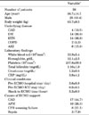
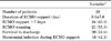
The mean patient age at the time of ECMO support was 56.7±14.7 years and 26 (68.4) patients were male. Among these, 14 (36.8%) patients had hypertension. Hospital and ICU stays before ECMO were 3.6±8.9 and 0.9±0.5 days, respectively. The duration of shock before ECMO initiation was 5.3±9.0 hours. Patient characteristics before ECMO support are summarized in Table 1. VA ECMO was the predominant type used in 92.1% (n=35) of patients. Veno-veno-arterial ECMO was used in 3 patients (7.9%). The duration of ECMO support was 9.0±7.6 days. Twenty-four (63.2%) patients received ECMO support for more than 7 days. There were 17 nosocomial infections in 16 (42.1%) patients. The rates of weaning success and survival to discharge were 55.3% (n=21) and 52.6% (n=20), respectively. The early outcomes of ECMO support are summarized in Table 2.
2. Statistical analysis of risk factors for survival
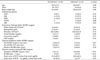
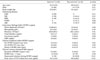
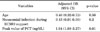
The baseline characteristics of 16 patients with infection and 22 without infection are shown in Table 3. There were no significant differences between the 2 groups in terms of sex, body weight, underlying disease, laboratory findings before ECMO support, ICU and hospital stay before ECMO, and shock to ECMO time. The duration of ECMO support was the only risk factor for occurrence of nosocomial infection during ECMO (p<0.01). However, the increase in PCT levels within the first week was not useful in predicting the occurrence of a new nosocomial infection during the ECMO run. The results of risk factor analysis for survival before and during ECMO support are summarized in Table 4. There were no significant differences between the 2 groups in terms of sex, body weight, underlying disease, laboratory findings before ECMO support, duration of ICU and hospital stay before ECMO, shock to ECMO time, and duration of ECMO support. The survivor group was younger than the non-survivor group (p=0.03). The non-survivor group had a higher incidence of nosocomial infection (p<0.01) and higher peak PCT levels (p<0.01). In logistic regression analysis, an independent predictor of survival before and during ECMO support was a higher peak PCT value (adjusted odds ratio, 1.94; 95% confidence interval, 1.03–3.27; p=0.01) (Table 5).
3. Association between survival outcome and PCT levels
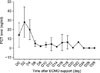
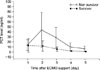
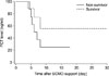
Fig. 2 shows the trends of PCT (mean values) during ECMO support. Maximal levels were reached within 48 hours after initiation of ECMO support (mean 25.6±9.4ng/mL), and decreased to ≤5 ng/mL within 1 week. The mean value of PCT levels after the first week varied in diverse patterns, but did not show the high levels shown for the first week. Serial progression of mean PCT levels was compared in the 2 groups during the first week of ECMO support; the mean PCT level was significantly higher in the survival group, according to time (p=0.03) (Fig. 3). In the Kaplan-Meier survival analysis, a PCT level of >10 ng/mL was significantly associated with survival probability within the first week of ECMO support (p=0.01) (Fig 4).
DISCUSSION
We evaluated the trends and predictive value of PCT levels during treatment of adult cardiogenic shock with ECMO, and investigated the role of PCT as a predictor of infection occurrence and clinical outcomes. As it is easy to measure and can be repeatedly checked, PCT has an important role as a predictor of infection. Therefore, many studies have used PCT as a guide to the use of antibiotics, and as a marker to monitor the effectiveness of antibiotic treatment in bacterial lower respiratory tract infections and sepsis.1213141516 PCT is also superior to traditional inflammatory markers such as CRP for diagnosis of bacterial infections.171819 In addition, higher PCT levels are associated with disease severity in septic patients.19 However, many recent studies have shown a negative correlation, suggesting that PCT measurement is not associated with decreased mortality or duration of antibiotic treatment in sepsis.112021
Studies on the usefulness of PCT measurement have been performed in various fields. Cardiopulmonary bypass (CPB), which is essential for cardiac surgery, causes a systemic inflammatory response, and the likelihood of an inflammatory reaction is increased when the running time is prolonged. As acute phase reactions following CPB are associated with organ dysfunction, there is a continuing effort to identify a biomarker for use in early diagnosis of complications after cardiac surgery. It is known that PCT affects the clinical course after cardiac surgery22 and is a more sensitive indicator than the increase in WBCs or CRP in predicting complications; moreover, the role of PCT is expanding to include prognostic use in specific acute cardiovascular diseases such as aortic dissection.23 Elevated PCT levels have a negative impact on postoperative complications and outcomes.1623 After cardiac surgery, PCT increases if the patient does not enter the recovery phase. Transfusion and deep hypothermia also induce a variety of inflammatory reactions, resulting in an increase in PCT levels during cardiac surgery.2425 Thus, it can be inferred that an elevation of PCT in the absence of specific symptoms can predict progressive deterioration. Therefore, PCT measurement after cardiac surgery can enable early detection and intervention to prevent complications. However, its value as a prognostic marker is still unclear due to a lack of evidence. To determine the mechanism by which PCT affects the body, a large-scale, randomized-control trial is needed to evaluate complications after cardiac surgery and to assess the value of other prognostic factors.
Although several studies have attempted to differentiate between increased PCT levels caused by infection versus ECMO, only a few preliminary studies have identified the role of PCT during ECMO therapy, and most have reported PCT as an indicator of infection.7826 One prospective study in pediatric patients found no correlation between PCT and prediction of infection, but did show that high PCT levels are associated with multi-organ dysfunction. 7 In adults, studies have identified the role of PCT as an indicator of infection during ECMO treatment.826 Our study showed that PCT was a potential serum biomarker for prognosis in patients undergoing ECMO support. Although our study could not precisely identify the change in PCT values correlated with disease severity, we confirmed that a peak value of >10 ng/mL was associated with mortality. However, when we divided patients into 2 groups based on a PCT level of 5 ng/mL, there was no statistically significant difference.
This study has some limitations. Firstly, this was a retrospective analysis of medical records. Thus, a selection bias was not completely excluded because 7.3% of cases were excluded, and association is not equivalent to causation. Second, this single-center study enrolled a relatively small number of patients. In the future, a large‑scale, prospective multicenter analysis is required to develop guidelines.
In conclusion, the level of PCT is not useful in prediction of nosocomial infection during ECMO support. However, higher PCT levels within the first week of ECMO are associated with significantly higher mortality.
 XML Download