

 PDF
PDF ePub
ePub Citation
 Print
Print


INTRODUCTION
Traumatic dental injury (TDI) is a public dental health problem because of a high frequency of occurrence, a high incidence during childhood, and associated treatment costs. Moreover, it often carries a lifelong treatment burden for the patient.1 Specific population studies have shown that trauma to the oral region can account for 5% of all injuries, with the highest risk in children aged 0 to 12 years.2 Guidelines for the management of traumatized teeth have been clearly outlined3; however, despite the dentist's best effort, tooth loss following trauma is sometimes inevitable. In order to select the best treatment option for patients with a lost anterior tooth, the clinician must consider several factors such as the patient's growth status, success rates and costs of various treatment options, patient preferences, and, most importantly, treatment options that will enable long-term rehabilitation.
Tooth autotransplantation to replace lost or missing teeth has been attempted for several centuries. Even early on, it became clear that there were two critically important factors for success. These included the use of an immature donor tooth for transplantation and the immediate transplantation of the immature tooth into the recipient site following extraction.4 Some of the early published works on autotransplantation appeared in the 1950s,56 followed by the case series by Slagsvold and Bjercke.7 Extensive animal studies and long-term prospective studies by Andreasen et al.8910 have served as stepping stones for the development of this treatment as a viable alternative for tooth substitution.
In a growing individual, the replacement of a lost anterior tooth by autotransplantation becomes a logical choice because it allows for continuous skeletal growth and the preservation of a vital periodontium.11 Dental implants, on the other hand, require long periods of space maintenance until the cessation of growth. Once the biological principles behind successful autotransplantation have been understood and the indications have been defined, it can be considered an extremely successful treatment modality with significant savings in time and cost compared with implants.12
Here we report the successful outcome of autotransplantation used in conjunction with orthodontic treatment for the replacement of a lost central incisor in a growing child who experienced trauma to the anterior maxilla. The uniqueness of this case is further emphasized by the unconventional decision to use the donor tooth from the arch that already had a congenitally missing tooth.
ETIOLOGY AND DIAGNOSIS
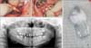
An 11-year-old boy was referred to the orthodontic clinic from the endodontic clinic for the interdisciplinary management of a traumatized anterior tooth. Dental history revealed a recent bicycle accident that resulted in trauma to the maxillary right central incisor (#11). Immediately after the injury was sustained, the patient was rushed to Pediatric Dentistry Clinic of University of Connecticut, where examination demonstrated a suspected coronal-radicular fracture as well as mobility of tooth #11. The maxillary anterior teeth were splinted with fiber-reinforced composite, and the patient was referred to the Endodontic clinic for further management. The splint was removed after 3 weeks, when clinical examination revealed a fracture line extending onto the labial and palatal surfaces of tooth #11 (Figure 1A and 1B). For further investigation, cone-beam computed tomography (CBCT) was performed, which confirmed the presence of a vertical crown fracture and also revealed a horizontal fracture at the cementoenamel junction/coronal portion of the root (Figure 1C). Because the prognosis of the fractured incisor was poor, the interdisciplinary team recommended the extraction of tooth #11.
TREATMENT OBJECTIVES
The primary objective was to determine a treatment plan that involved temporary or permanent replacement of tooth #11, which was lost because of trauma, as well as the restoration of esthetics in the maxillary anterior region. The secondary objective was to maintain the maxillary incisor inclination and/or obtain good functional occlusion.
TREATMENT ALTERNATIVES
The following treatment alternatives were considered.
Dental implantation for the replacement of tooth #11. This would involve initial orthodontic treatment to manage the space of tooth #11 and position the adjacent tooth roots ideally for the future implant and fixed retention across the edentulous space to prevent tipping of the adjacent roots, along with a temporary pontic for tooth #11 for esthetics and space maintenance.
Autotransplantation of a mandibular premolar (#45) into the region of the extracted tooth, followed by orthodontic treatment to close all spaces and obtain good functional occlusion. Restorative and esthetic recontouring of the autotransplanted tooth would be performed at the end of orthodontic treatment.
The patient's parents and the interdisciplinary team opted for the second alternative, because it was considered to provide the best long-term prognosis and would consequently prove the most beneficial for the patient.
TREATMENT PROGRESS
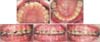
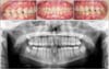
The fractured central incisor was extracted and the recipient site was prepared to receive the donor tooth. Subsequently, tooth #45 was atraumatically extracted, placed in the recipient site, and splinted with a 0.020-inch stainless steel wire that was bent passively to fit the labial contour of the tooth and was extended to be engaged into the bracket slots on the adjacent maxillary incisors (Figure 2A and 2B). The patient reported to the orthodontic clinic 4 weeks after the autotransplantation procedure. At that time, orthodontic examinations and records were completed. Intraorally, the patient was in the late mixed dentition stage with a congenitally missing mandibular central incisor, Class I molar relationship, and normal overjet and overbite. The panoramic radiograph showed alveolar bone remodeling around the autotransplanted tooth and the edentulous space of the donor tooth (Figure 2C). Cephalometric analysis showed a mild Class III skeletal relationship (A point, nasion, B point [ANB] = 0°), an upper incisor inclination of 105°, and a nasolabial angle of 104°.
Specific orthodontic treatment objectives involved closure of the donor tooth space and movement of the autotransplanted tooth into the ideal position so that it could be restored to achieve the best esthetic outcome. The final plan involved the orthodontic closure of all spaces to eliminate the need for future dental implants, followed by restorative build-ups.
Three months after autotransplantation, 0.022-inch MBT™ prescription metal brackets were bonded on the maxillary and mandibular teeth and initial archwires were placed. To achieve absolute protraction of the mandibular first molar, which was required to achieve a Class III functional occlusion, a mini-implant (2.0 × 9 mm Lomas; Mondeal Medical Systems GmBH, Donau, Germany) was placed on the alveolar ridge distal to the mandibular right first premolar (Figure 3).
RESULTS
The total treatment duration was 36 months. The autotransplanted tooth was esthetically restored during the finishing phase of the orthodontic treatment; this also helped in achieving good functional occlusion at the end of the orthodontic treatment (Figure 4A). The patient and his parents were pleased with the results, considering the interdisciplinary approach had resulted in the esthetically pleasing replacement of the lost central incisor. The post-treatment panoramic radiograph showed that the root formation had progressed to achieve a full length (Figure 4B). Post-orthodontic evaluation of the autotransplanted tooth demonstrated all signs of a successful transplant,13 which include a good crown to root ratio, no external root resorption or mobility, and, most importantly, an appearance that is identical to the counterpart, with a matching gingival contour and emergence profile.
DISCUSSION
We reported the successful outcome of autotransplantation used in conjunction with orthodontic treatment for the replacement of a lost central incisor in a growing child who experienced trauma to the anterior maxilla. The novelty of our case is that, autotransplantation was done in a patient who did not meet the typical criteria. Traditionally, autotransplantion is considered, when extraction in one of the arches is indicated as part of orthodontic treatment planning; as in, there is requirement for additional space so as to relieve crowding or address proclined teeth. Our case demonstrates that, even if the strictest criteria for autotransplantation are not met, it should not deter the orthodontic professionals from wanting to use this option as we are in a generation where we can use tools like three-dimensional CBCT and temporary anchorage devices to ensure a more predictable outcomes.
The treatment of TDI in a young patient is often unpredictable, complicated, expensive, and can continue throughout his or her life; therefore, the treatment of choice should minimize these undesired consequences.14 The treatment options include removable or conventional fixed prostheses, dental implant-supported crowns, autotransplantation, and simple orthodontic space closure with tooth substitution.15
Currently, an implant-supported restoration is considered the gold standard for the conservative replacement of a lost tooth in adult patients.15 In a growing child, this treatment modality generally necessitates space maintenance until facial growth is completed. During this time, there is continued alveolar bone remodeling in terms of both width and height; this may require alveolar bone grafting procedures and consequently increase the total cost of treatment. Furthermore, a long-term study demonstrated that the cessation of vertical growth is not very predictable, with mature adults exhibiting vertical steps after the placement of anterior implant restorations to the same extent as patients in the late adolescent stage.16 Autotransplantation of teeth can be an excellent treatment alternative to overcome the above concerns in young adolescents. The successful transplantation of a premolar along with healthy surrounding tissues in our patient confirms that this treatment modality can be recommended for the replacement of missing maxillary incisors in young patients.
Successful tooth autotransplantation results in the maintenance of a viable periodontal ligament and, consequently, the potential for bone induction and the capacity for continuous eruption during growth, leading to the re-establishment of a normal alveolar process.11 Because autotransplantation is a technique- and operator-sensitive procedure, it has not been established as a standard-of-care for the replacement of lost teeth. This may also be the reason for the considerably varied success and survival rates for autotransplanted teeth reported in the literature. An overall comparison of the survival rates for autotransplanted teeth with those for single-tooth osseointegrated implants demonstrated comparable results. Czochrowska et al.17 showed a 90% survival rate for autotransplanted teeth over a follow-up period of 17 to 41 years and a 94.5% survival rate for single-tooth implants over a period of 5 years.18

 XML Download