

 PDF
PDF ePub
ePub Citation
 Print
Print


INTRODUCTION
The structure and function of bone is regulated through bone remodeling, which occurs in microscopic basic multicellular units (BMU) that consist of bone-forming osteoblasts, bone-resorbing osteoclasts, and osteocytes within the bone matrix, the bone lining cells that cover the bone surface and the capillary blood supply [12].
The bone remodeling cycle is composed of three phases: initiation, reversal, and termination. The initiation phase includes the migration of osteoclast precursors to the bone surface, their differentiation into mature osteoclasts, and the maintenance of bone resorption. The reversal phase is a transition from osteoclast to osteoblast activity. The final stage is the termination phase, in which bone formation by osteoblasts occurs [34]. An imbalance in bone remodeling can result in an excess of bone resorption compared with bone formation, which may lead to the occurrence of pathologic states of bone structure, such as osteoporosis.
Osteoclasts are a key participant in bone remodeling. Located on endosteal surfaces within the Haversian system and on the periosteal surface beneath the periosteum, these are multinucleated, giant cells formed by the fusion of mononuclear progenitors of the monocyte/macrophage family by osteoclastogenesis [56]. For osteoclastogenesis, macrophage colony-stimulating factor (M-CSF) and receptor for activation of nuclear factor kappa B ligand (RANKL) are required. These molecules induce the expression of osteoclastogenic genes, such as tartrate-resistance acid phosphatase (TRAP), cathepsin K (CTK), calcitonin receptor (CTR), and β3-integrin, which lead to the formation of mature osteoclasts [7]. Cell fusion in the osteoclast lineage is necessary for the formation of mature osteoclasts (also known as multinucleated osteoclasts). The main effect of osteoclast multinucleation is an increase in the size of osteoclasts, which causes their activation and stimulates the resorption of larger bone tissue [89]. Therefore, osteoclastogenic cell fusion is an essential component of the process of osteoclastogenesis. Many molecules involved in osteoclastogenic cell fusion have been reported, including macrophage fusion receptor, signal regulatory protein α, and dendritic cell-specific transmembrane protein (DC-STAMP) [1011].
Remifentanil is a short-acting µ-opioid receptor agonist that is rapidly hydrolyzed by nonspecific esterases in plasma and tissue; even when administered as a prolonged infusion, it does not accumulate. Therefore, it can be administered via continuous infusion during general anesthesia and sedation in the intensive care unit (ICU) [1213]. During orthognathic surgery or prolonged sedation in ICU, the continuous infusion of remifentanil may affect the bone physiology of the patient, because the patients who undergo orthognathic surgery or are treated in the ICU are more likely to have pathologic bone disease or to be elderly. The effects of remifentanil on osteoblasts have been previously studied. It was recently reported that remifentanil has a protective effect against oxidative stress on human osteoblasts [14]. Baik et al. [15] determined that remifentanil preconditioning enhanced the maturation of osteoblasts under hypoxia- reoxygenation conditions and increased the expression of osteoblastogenic genes. However, the effects of remifentanil on osteoclast differentiation have not been reported. Therefore, we investigated the effects of remifentanil on osteoclast differentiation and osteoclastogenic gene expression through the in vitro examination of pre-osteoclasts.
MATERIALS AND METHODS
1. Reagents
Remifentanil was obtained from GlaxoSmithKline Pharmaceuticals (Rockville, MD, USA) and dissolved in research-grade deionized distilled water (DNAse- and RNAse-free). M-CSF and recombinant RANKL were purchased from PeproTech (Rocky Hill, NJ, USA). Antibodies against c-fos and nuclear factor of activated T cell cytoplasmic 1 (NFATc1) were obtained from SantaCruz Biotechnology (Santa Cruz, CA, USA). The Leukocyte Acid Phosphatase (TRAP) kit and anti-β-actin antibody were obtained from Sigma-Aldrich (St. Louis, MO, USA). All other chemicals and reagents were purchased from Sigma-Aldrich.
2. Isolation of bone marrow-derived macrophages (BMMs) and in vitro osteoclastogenic differentiation
BMMs were isolated from mice bone marrow and used as osteoclast precursor cells for in vitro osteoclast differentiation, as previously described [16]. Mouse whole bone marrow cells were collected from the femurs and tibiae of 5-week-old ICR mice, placed in 100 mm dishes, and cultured overnight in α-modified Eagle Medium (α-MEM; WelGENE Inc., Daegu, Korea) supplemented with 10% fetal bovine serum (FBS). Adherent stromal cells were discarded and the floating cells were cultured in the presence of M-CSF (30 ng/ml) in Petri dishes. The cells became adherent after 3–4 days of culture and were used as BMMs, the osteoclast precursor cells. Animal experiments were approved by the Committees on the Care and Use of Animals in Research at Pusan National University. To induce osteoclast differentiation, BMMs (4 × 10⁴ cells/48-well plates) were cultured with osteoclastogenic medium (OM: 30 ng/ml M-CSF + 100 ng/ml RANKL) for 4 days. After culture, the cells were stained for TRAP activity and the TRAP-positive multinucleated cells with ≥ 3 nuclei were considered to be osteoclasts.
3. Pre-osteoclast (pre-OC) differentiation and remifentanil treatment
The differentiation of pre-OCs, mononuclear TRAP-positive cells, was induced through the culture of BMMs with OM for 2 days. The pre-OCs were treated with fresh α-MEM (with M-CSF and RANKL) in the presence or absence of various concentrations of remifentanil and cultured for up to 3 days (5 days in total). All experiments were conducted on pre-OCs.
Cytotoxicity measurement and cell proliferation assay The effects of remifentanil on both cell viability and proliferation were examined by the well-established colorimetric 3-(4,5-dimethylthiazol)-2,5-diphenyltetrazolium bromide (MTT, Sigma-Aldrich) assay. In brief, pre-OCs were seeded in 96-well plates with the indicated concentrations of remifentanil (0.1, 1, 10, and 100 ng/ml) for up to 3 days. At the end of the culture period, the cells were incubated with MTT solution (medium containing 0.5 mg/ml MTT) for a further 4 h. After culture, the formation of the blue formazan product was measured at 570 nm by using a microplate reader.
4. TRAP staining
The cells were fixed in 3.7% formalin for 10 min, washed twice with phosphate buffered saline (PBS), and permeabilized by the addition of 0.3 mg/ml Fast Red Violet LB in 0.05 M sodium acetate, 0.05 M acetic acid, 0.03 M sodium tartrate, 0.1 mg/ml naphthol AS-MX phosphate disodium salt, and 0.1% Triton X-100 for 1 min. The cells were the washed with PBS and stained for TRAP activity by using a TRAP kit (Sigma) in accordance with the manufacturer's instructions. After incubation at 37℃ for 1 h, the cells were washed three times with distilled water. TRAP-positive cells appeared red or purple.
5. Real-time polymerase-chain reaction (PCR) analysis
Total RNA was isolated by using TRIzol reagent (Invitrogen, CA, USA) and 1.5 µg was reverse-transcribed by using Superscript II (Invitrogen) in accordance with the manufacturer's protocol. For real-time PCR analysis, 2 µg of cDNA was amplified with SYBR green PCR master mix (Applied Biosystems) for 40 cycles of 15 s at 95℃ for denaturation and 1 min at 60℃ for amplification by using an AB7500 (Applied Biosystems). Relative mRNA expression levels were obtained after normalizing against actin mRNA levels. The primer sets used in real-time PCR were as follows: NFATc1, 5′-CCA GTA TAC CAG CTC TGC CA-3′ (forward) and 5′-GTG GGA AGT CAG AAG TGG GT-3′ (reverse); c-fos, 5′-ACT TCT TGT TTC CGG C-3′ (forward) and 5′-AGC TTC AGG GTA GGT G-3′ (reverse); DC-STAMP, 5′-GGG TGC TGT TTG CCG CTG-3′ (forward) and 5′-CGA CTC CTT GGG TTC CTT GCT-3′ (reverse); CTK, 5′-ATA TGT GGG CCA CCA TGA AAG TT-3′ (forward) and 5′-TCG TTC CCC ACA GGA ATC TCT-3′ (reverse); CTR, 5′-GCA ACC GAA CCT GGT CCA ACT AT-3′ (forward) and 5′-AAG CAG CAA TCG ACA AGG AGT GA-3′ (reverse); Actin, 5′-TCT GGC ACC ACA CCT TCT AC-3′ (forward) and 5′-TAC GAC CAG AGG CAT ACA GG-3′ (reverse).
6. Boyden chamber migration assay
Transwells with 8 µm porosity polycarbonate membrane inserts were used. Pre-OCs were suspended in α-MEM media at 1×10⁵ cells/100 µl and were added to the upper chamber. Remifentanil (10 and 50 ng/ml) in α-MEM media was added into the lower chamber. Pre-OCs that migrated through the filter and appeared on the lower side were fixed by careful immersion of the filter into methanol for 1 min, stained with hematoxylin and eosin (H&E) solution, and counted in three random fields per well. Each experiment was performed in duplicate and three separate experiments were performed for each treatment group.
7. Statistical analysis
Data were obtained from at least three independent experiments. Student's t-test was used to determine the significance of differences between two groups. Differences with P < 0.01 were regarded as statistically significant and denoted with an asterisk (**P < 0.01; ***P < 0.001).
RESULTS
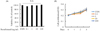
1. Remifentanil was not cytotoxic to pre-OCs and did not increase cell proliferation
BMMs were cultured with OM for 2 days to induce differentiation to pre-OCs (Fig. 1A). The effect of remifentanil on the cell viability and proliferation of pre-OCs was verified by using an MTT assay. The MTT assay indicated that the cell viability of pre-OCs was not affected by treatment with various concentrations of remifentanil (0.1, 1, 10, and 100 ng/ml) (Fig. 1B). The proliferation of pre-OCs cultured in OM for 3 days with remifentanil was maintained and no significant difference in cell proliferation was identified at different remifentanil concentrations (Fig. 1B).
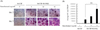
2. Remifentanil increased pre-OC differentiation and the size of osteoclasts cultured with M-CSF and RANKL
Pre-OCs were cultured with M-CSF alone or OM in the presence of remifentanil for 2–3 days to evaluate the formation of mature osteoclasts. TRAP staining was used for this analysis; TRAP-positive multinucleated cells with ≥ 3 nuclei were considered to be mature osteoclasts. Remifentanil increased the formation of TRAP-positive multinucleated osteoclasts, especially after 3 days (Fig. 2A). The size of osteoclasts was significantly increased by the presence of remifentanil (50 ng/ml) compared with the absence of remifentanil (Fig. 2B). These results confirmed that the administration of remifentanil to pre-OCs enhanced osteoclast differentiation and maturation.
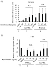
3. The mRNA expression of NFATc1 and c-fos was not affected by remifentanil treatment
NFATc1, a member of the NFAT family of transcription factor genes [17], is known as key transcription factor that induces a number of genes involved in osteoclast differentiation. It is strongly induced following RANKL stimulation. The induction of NFATc1 following RANKL is regulated by intracellular calcium oscillation and another transcription factor, c-fos [1718]. To investigate the effect of remifentanil on the expression of transcription factors such as NFATc1 and c-fos, the relative mRNA expression of NFATc1 and c-fos genes from pre-OCs treated with M-CSF alone or M-CSF and RANKL for 2 and 4 days was measured by using quantitative real-time PCR. The relative mRNA expression of NFATc1 and c-fos in pre-OCs was not altered by an increase in the dose of remifentanil (0, 10, and 50 ng/ml) during osteoclastogenesis (Fig. 3A, B). These results confirmed that the enhancement of pre-OC differentiation by remifentanil was not mediated by the activation of NFATc1 and c-fos.
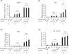
4. Remifentanil did not induce the expression of osteoclast-specific genes such as TRAP, CTK, CTR, and DC-STAMP
TRAP, CTK, and CTR are osteoclast differentiation marker genes that are induced by NFATc1 activation at the final stage of differentiation [19]. As previous results showed that remifentanil upregulated pre-OC differentiation (Fig. 2A, B), we hypothesized that remifentanil may increase the expression of osteoclast-specific genes. The mRNA expression of osteoclastogenic-specific genes was analyzed in pre-OCs. As shown in Fig. 4A, B, and C, the relative mRNA expression of TRAP, CTK, and CTR was not significantly increased for remifentanil treatment (10 and 50 ng/ml) compared with the presence of only M-CSF and RANKL on days 2 and 4.
DC-STAMP is a key regulator in osteoclast cell-cell fusion, which is a critical process for osteoclast maturation [20]. To investigate the relationship of cell-cell fusion with the upregulation of pre-OC differentiation induced by remifentanil, the mRNA expression of DC-STAMP was evaluated. Treatment with remifentanil increased the relative mRNA expression of DC-STAMP in a dose-dependent manner after 4 days, but the difference was not significant (Fig. 4D).
5. Remifentanil enhanced pre-OC migration
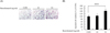
The migration of pre-OCs was evaluated by using the Boyden chamber assay. Pre-OCs in medium were seeded in the upper part of a Transwell plate and allowed to migrate vertically through the pores of the membrane into the lower chamber, which held the control or remifentanil-containing medium. The number of migrated cells was increased after remifentanil treatment (10 and 50 ng/ml) in the lower chamber compared with the control (Fig. 5A). In addition, the number of migrated cells was significantly increased by 50 ng/ml remifentanil compared with the untreated control (Fig. 5B).
DISCUSSION
In this study, we evaluated the effects of remifentanil on pre-OC differentiation and the expression of osteoclastogenic genes and transcription factors. The main findings of this study were: (1) remifentanil increased pre-OC differentiation and the size of osteoclasts in the presence of M-CSF and RANKL; (2) the increase of pre-OC differentiation induced by remifentanil was not related to the expression of the osteoclast-specific genes TRAP, CTK, CTR, and DC-STAMP or the expression of the osteoclastogenic transcription factors NFATc1 and c-fos; (3) the increase in the size of osteoclasts induced by remifentanil was mediated by the upregulation of osteoclast cell migration and subsequent cell fusion.
Osteoclast differentiation requires a number of osteoclastogenic factors for the regulation of these processes. M-CSF, produced by osteoblasts and stromal cells, is required for osteoclast precursor cell survival and activates early transcription factors, such as c-fos and PU. 1 [2122]. RANKL, expressed by osteoblasts, T cells, and endothelial cells, is indispensable for osteoclast formation [7]. The RANKL signaling pathway is a key pathway in the formation and function of osteoclasts. When RANKL binds to receptor activator of nuclear factor kappa B (NF-κB), it is translocated to the nucleus for the transcription of essential osteoclast genes. The RANKL pathway cooperates with the immunoreceptor tyrosine-based activation motif (ITAM) co-stimulatory pathway to activate calcium/NFATc1 signaling, which is necessary for osteoclastogenesis [23]. Thus, there are many signaling pathways and osteoclastogenic factors involved in osteoclast differentiation. In this study, remifentanil increased pre-OC differentiation, but did not increase the expression of the osteoclastogenic transcription factors, NFATc1 and c-fos. These results suggested the increase in pre-OC differentiation induced by remifentanil may be mediated through factors other than NFATc1 and c-fos.
Cell fusion is a characteristic properties of osteoclasts. Through this process, osteoclasts become multinucleated, increase in size, and are able to absorb bone [8]. In this study, the mRNA expression of DC-STAMP, a key protein for cell fusion in osteoclast, was not upregulated by remifentanil. This suggested that the increase in size of osteoclasts induced by remifentanil was not mediated by the upregulation of DC-STAMP. Cell fusion also can be enhanced by cell migration [24]. Therefore, we assessed the effect of remifentanil on pre-OC migration by using a Boyden chamber assay. The results showed that remifentanil enhanced pre-OC migration. These findings suggested pre-OC migration enhanced cell fusion, which resulted in an increase in osteoclast size. In a previous in vitro study, remifentanil suppressed the RANKL-induced osteoclast differentiation (data not shown). In this study, by contrast, remifentanil treatment promoted osteoclast differentiation. The difference between the two studies was the stage of differentiated osteoclasts at the time of remifentanil treatment. During osteoclastogenesis, hematopoietic precursor cells differentiate to mature osteoclast via several steps. First, BMMs differentiate to mononucleated osteoclasts (pre-OCs). Thereafter, cell-cell fusion of pre-OCs occurs and multinucleated osteoclasts (mature osteoclasts) are formed [25]. In previous study, the effects of remifentanil on osteoclast differentiation were investigated in BMMs. However, in this study, we added remifentanil to pre-OCs and evaluated osteoclast differentiation and other parameters. Contrasting results of osteoclast differentiation have been reported. A recent report stated that lipopolysaccharide (LPS) promoted osteoclast differentiation; in this study, LPS was added during the process of osteoclast differentiation after M-CSF and RANKL treatment [26]. In another study, LPS treatment to BMMs inhibited RANKL activity and subsequently decreased osteoclast differentiation [27]. Therefore, the effects of remifentanil on osteoclast differentiation may differ according to the stage of the differentiated osteoclasts treated with remifentanil.
In summary, we found that remifentanil increased pre-OC differentiation and the size of osteoclasts. In addition, we demonstrated that the remifentanil-induced increase in osteoclasts was mediated through the enhancement of pre-OC migration and cell fusion.
 XML Download