

This article has been corrected. See "Korean Clinical Imaging Guideline for Hemoptysis" in Volume 78 on page 299.
 PDF
PDF ePub
ePub Citation
 Print
Print


Abstract
In 2014, the American College of Radiology announced a guideline for appropriate diagnostic approach and treatment in patients with hemoptysis, according to severity of hemoptysis and risk of lung cancer. However, in Korea many patients have pulmonary fibrosis due to previous tuberculosis or have active tuberculosis. Therefore, application of this guideline is not appropriate. The Korean Society of Radiology and Korean Society of Thoracic Radiology proposed a guideline more closely matching the real state of diagnostic approach and treatment of patients with hemoptysis in Korea. The guideline was prepared in consensus by a development committee, working party, and an advisory committee. The process of the guideline proposal was based on methodology for developing evidence-based clinical imaging guidelines: joint recommendations by the Korean Society of Radiology and National Evidence-Based Healthcare Collaborating Agency. The clinical imaging guideline for adult patients with hemoptysis is as follows. Chest radiography is an initial imaging modality to evaluate hemoptysis. Contrast enhanced chest CT is recommended in patients with two lung cancer risks (> 40 years old and > 30 packs per year smoking history), moderate hemoptysis (> 30 cc/24 hours) or recurrent hemoptysis. Contrast enhanced chest CT is also recommended for patients with massive hemoptysis (> 400 mL/24 hours) without cardiopulmonary compromise.
Go to : 
References
1. ACR Appropriateness Criteria® hemoptysis. Available at:. https://acsearch.acr.org/docs/69449/Narrative/. Published. Aug, 2010. Accessed Jan 31, 2017.
2. Choi SJ, Jeong WK, Jo AJ, Choi JA, Kim MJ, Lee M, et al. Methodology for developing evidence-based clinical imaging guidelines: joint recommendations by Korean Society of Radiology and National Evidence-Based Healthcare Collaborating Agency. Korean J Radiol. 2017; 18:208–216.

3. Tsoumakidou M, Chrysofakis G, Tsiligianni I, Maltezakis G, Siafakas NM, Tzanakis N. A prospective analysis of 184 hemoptysis cases: diagnostic impact of chest X-ray, computed tomography, bronchoscopy. Respiration. 2006; 73:808–814.
4. Fidan A, Ozdog˘an S, Oruç O, Salepçi B, Ocal Z, Cag˘layan B. Hemoptysis: a retrospective analysis of 108 cases. Respir Med. 2002; 96:677–680.
5. Bruzzi JF, Rémy-Jardin M, Delhaye D, Teisseire A, Khalil C, Rémy J. Multi-detector row CT of hemoptysis. Radiographics. 2006; 26:3–22.
6. Lee SJ, Rho JY, Yoo SM, Kim MD, Lee JH, Kim EK, et al. Usefulness of multi-detector computed tomography before bronchoscopy and/or bronchial arterial embolization for hemoptysis. Tuberc Respir Dis. 2010; 68:80–86.
7. Revel MP, Fournier LS, Hennebicque AS, Cuenod CA, Meyer G, Reynaud P, et al. Can CT replace bronchoscopy in the detection of the site and cause of bleeding in patients with large or massive hemoptysis? AJR Am J Roentgenol. 2002; 179:1217–1224.
8. Delage A, Tillie-Leblond I, Cavestri B, Wallaert B, Marquette CH. Cryptogenic hemoptysis in chronic obstructive pulmonary disease: characteristics and outcome. Respiration. 2010; 80:387–392.
9. Menchini L, Remy-Jardin M, Faivre JB, Copin MC, Ramon P, Matran R, et al. Cryptogenic haemoptysis in smokers: angiography and results of embolisation in 35 patients. Eur Respir J. 2009; 34:1031–1039.
10. Poe RH, Israel RH, Marin MG, Ortiz CR, Dale RC, Wahl GW, et al. Utility of fiberoptic bronchoscopy in patients with hemoptysis and a nonlocalizing chest roentgenogram. Chest. 1988; 93:70–75.
11. Herth F, Ernst A, Becker HD. Long-term outcome and lung cancer incidence in patients with hemoptysis of unknown origin. Chest. 2001; 120:1592–1594.
12. Thirumaran M, Sundar R, Sutcliffe IM, Currie DC. Is investigation of patients with haemoptysis and normal chest radiograph justified? Thorax. 2009; 64:854–856.
13. McGuinness G, Beacher JR, Harkin TJ, Garay SM, Rom WN, Naidich DP. Hemoptysis: prospective high-resolution CT/bronchoscopic correlation. Chest. 1994; 105:1155–1162.
14. Kim HB. Bronchial artery embolization. Korean Society of Interventional Radiology. ed.Interventional radiology. 2nd ed.Seoul: Ilchokak;2014. p. 321–325.
15. Millar AB, Boothroyd AE, Edwards D, Hetzel MR. The role of computed tomography (CT) in the investigation of unexplained haemoptysis. Respir Med. 1992; 86:39–44.
16. Khalil A, Fartoukh M, Parrot A, Bazelly B, Marsault C, Carette MF. Impact of MDCT angiography on the management of patients with hemoptysis. AJR Am J Roentgenol. 2010; 195:772–778.
17. Hsiao EI, Kirsch CM, Kagawa FT, Wehner JH, Jensen WA, Baxter RB. Utility of fiberoptic bronchoscopy before bronchial artery embolization for massive hemoptysis. AJR Am J Roentgenol. 2001; 177:861–867.
Go to :
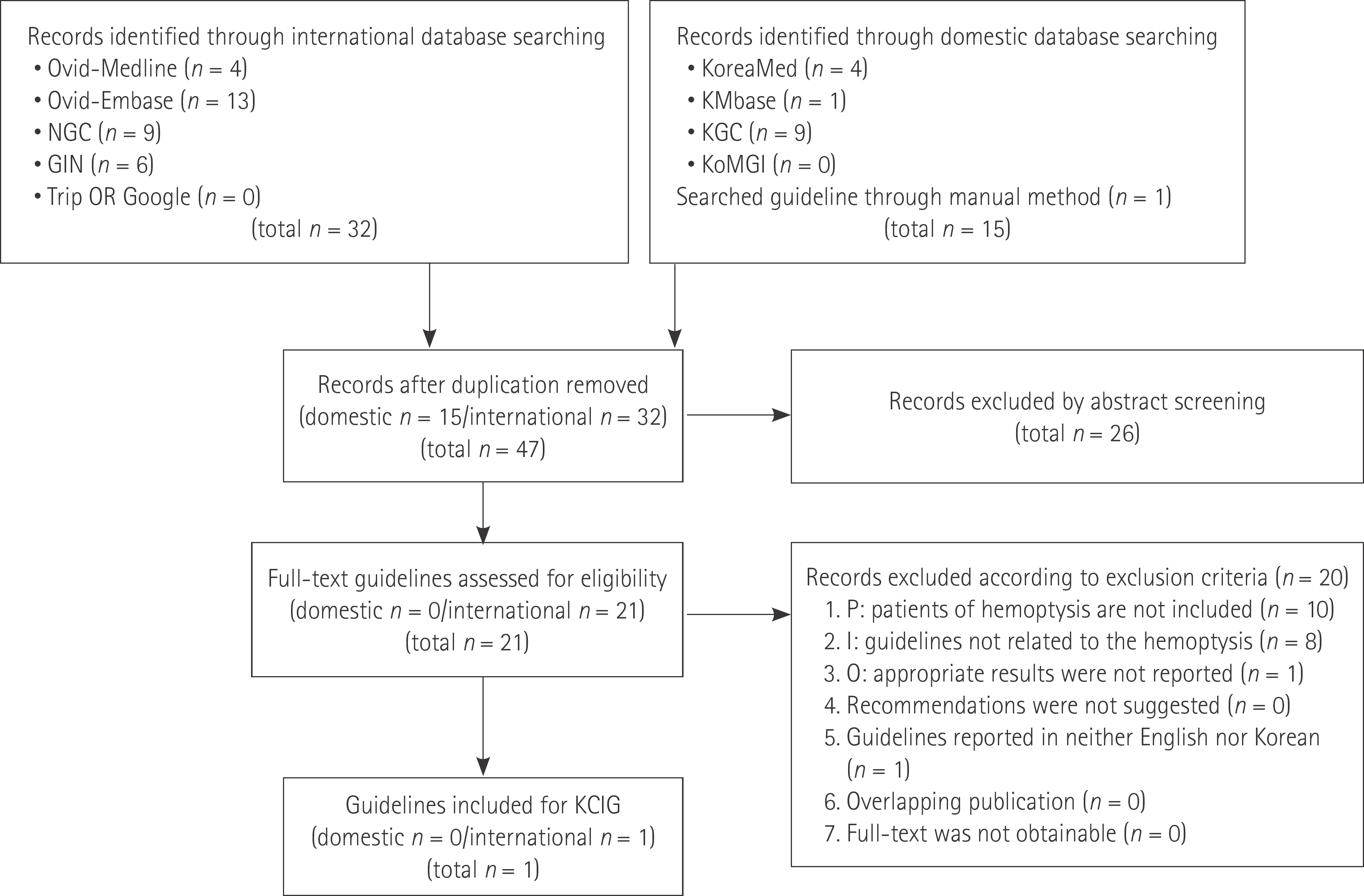 | Fig. 1.Flow chart for literature selection. GIN = Guideline International Network, I = intervention/index test, KCIG = Korean Clinical Imaging Guidelines, NGC = National Guideline Clearinghouse, O = outcome, P = population/patient |
Table 1.
AGREE II
| Source of Recommendation | AGREE II Score | Proposal of Develomental Committee |
|---|---|---|
| ACR Appropriateness Criteria® hemoptysis. | 66 | Recommended |
Table 2.
Criteria for Evidence Level of Each Evidence Literatures
Table 3.
Grades of Korean Clinical Imaging Guidelines Recommendation
Supplementary Table 1
Evidence Table
 XML Download