

 PDF
PDF ePub
ePub Citation
 Print
Print


INTRODUCTION
Lymphedema is characterized by the collection of excessive protein-rich fluid in the regional interstitium. It results from abnormalities such as lymphatic hypoplasia, functional insufficiency or absence of lymphatic valves, and lymphatic stasis in regional lymphatic drainage of the extremities (1). These abnormalities allow the build-up of protein and fluid within the interstitium, resulting in edema, pain, and skin changes.
Clinically, a diagnosis of lymphedema is made after excluding other pathologic conditions such as chronic venous insufficiency, post-phlebitic syndrome, myxedema, and lipedema (1). After excluding these conditions, several causes of lymphedema such as an inherent defect in the lymph vessels or nodes; surgical, traumatic, inflammatory, or neoplastic disruption; and obstruction of lymphatic pathways should be considered (1). The most common causes of lymphedema are surgical trauma such as lymph node dissection and radiation therapy (2). Additionally, although it rarely occurs, critical traumatic injury such as extensive burns and massive fractures can cause lymphedema, as described in previous case reports (3456789).
Here, we report a case wherein the patient had a swollen leg resulting from only a minor trauma and this showed abnormal findings on lymphoscintigraphy, suggesting lymphedema. As lymphedema induced by minor trauma has not been reported yet, this is the first report to suggest that minor trauma can cause lymphedema and that lymphoscintigraphy can provide critical information for its diagnosis.
CASE REPORT
A 72-year-old man visited our hospital for severe right lower limb swelling after right ankle contusion caused by falling off a ladder 8 months ago. Physical exams and simple radiography performed at the time of the accident showed no limitation in the range of motion or visible fractures. After the accident, the swelling developed leading to an increased circumference in the leg. He also had a history of taking anti-tuberculosis medication and had undergone surgery for abdominal tuberculous lymphadenitis approximately 50 years ago, without reactivation after the treatment.
First, we investigated the systemic causes of limb swelling, including congestive heart failure, renal failure, and hypoalbuminemia. Blood tests revealed mildly decreased levels of protein (4.2 g/dL; reference value 5.8–8.1 g/dL) and albumin (2.8 g/dL; reference value 3.1–5.2 g/dL). Blood urea nitrogen (16.4 mg/dL; reference value 8–20 mg/dL) and creatinine levels (0.8 mg/dL; reference value 0.5–1.2 mg/dL) were within normal range. There was no abnormal finding in the urinalysis. Cardiac evaluation including echocardiography also showed no abnormal findings.
Following this, we performed diagnostic examinations for local causes such as deep vein thrombosis, infections, lipedema, lymphedema, and malignancies. The patient gave written consent for all tests. Lower extremity computed tomographic venography showed a marked expansion of the subcutaneous fat layer and fibrosis in the right truncal and lower leg area without visible thrombosis. Contrast-enhanced abdominopelvic computed tomography (CT) images revealed multiple lymph nodes with internal calcifications without evidence of any malignancy or infectious disease. A chest CT showed only old sequelae of tuberculosis without evidence of reactivation.
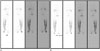
As other diagnostic examinations showed no significant abnormalities, lymphoscintigraphy was performed using Tc-99m phytate to assess the lymphatic flow. Tc-99m phytate was injected subcutaneously in the interdigital web spaces of both feet. The image, which was obtained at one and two hours after the injection, showed delayed lymphatic flow and dermal backflow with collateral lymphatics in the whole right lower limb (Fig. 1). In addition, the right inguinal lymph node showed no radiotracer uptake, suggesting lymphatic flow obstruction below the inguinal lymph node level.
When a detailed history of the patient was taken, the clinician found that the patient had mild pain in the right inner thigh as a result of falling off a ladder 8 months ago. As there was no tissue injury or fracture at that time, the patient assumed that the pain was the result of contusion injury, and the pain resolved after a few days. Considering the findings of the lymphoscintigraphy and previous history, the clinician diagnosed lymphatic obstruction, caused by minor traumatic injury.
In this situation, surgical correction of lymphatic flow was the treatment of choice. However, the patient was too old to undergo surgical treatment; hence, the clinician prescribed conservative treatments such as lymph massage and compression stockings. As the circumference of the right lower limb has improved after the treatment, the patient was discharged.
DISCUSSION
This is the first case of secondary lymphedema induced by minor trauma and detected by lymphoscintigraphy. After the occurrence of minor trauma, progressive edema of the right lower leg developed in the patient. On diagnostic examinations, only lymphoscintigraphy showed abnormal findings suggestive of lymphatic obstruction below the level of the inguinal lymph node with diffusely developed collateral lymphatic flows. Medical history and lymphoscintigraphy indicated that one minor injury could cause severe and progressive lymphedema trauma even when there was no internal wound.
Lymphedema is one of the local causes of limb swelling (8). The most common cause of secondary lymphedema in developed countries is iatrogenic, a direct consequence of surgical and radiotherapeutic interventions for malignancy (3). Lymphedema can also result from other forms of trauma such as burn injuries and severe damage to the extremity (3). Some authors have shown that secondary lymphedema can be caused by trauma as the local lymph nodes can react to internal wounds such as bone fractures and injury to adjacent tissues through the mobilization of cells from blood circulation along with the activation of cellular subsets (10). However, in the present case, the patient only had a history of minor trauma without any sign of injury to the soft tissue or bony structure. Previous reports have described lymphedema only resulting from severe trauma in the limbs (45679).
It is difficult to treat lymphedema once it progresses; therefore, early diagnosis with proper treatment is important. Ultrasound and duplex ultrasound can evaluate volumetric and structural changes as well as gradual impedance of venous return (8). However, the techniques show limitations in evaluating truncal anatomy of the lymphatics (8). CT and magnetic resonance imaging can help diagnose lymphedema by demonstrating thickening of skin and subcutaneous tissue with a typical honeycomb appearance (8). However, their diagnostic value is limited when calf swelling is not present. In addition, they cannot distinguish primary lymphedema from secondary causes (8). For evaluation of the anatomy of lymphatics, lymphangiography and lymphoscintigraphy are the gold standard (8). They can measure lymphatic function, lymph movement, lymph drainage, and response to treatment of lymphedema (8). However, lymphangiography is an invasive procedure that may lead to infection, local inflammation, and fibrosis (8). In contrast, lymphoscintigraphy is non-invasive, and is the most sensitive tool for visualizing lymphatic obstruction (10). Lymphoscintigraphy is particularly useful in diagnosing lymphatic dysfunction in a swollen limb. With abnormal findings of lymphoscintigraphy including delayed, cut-off, collateral lymphatic flow; dermal backflow; and non-visualization of lymph node uptake, a diagnosis of lymphatic obstruction can be made.
In the present case, the patient had a progressively swollen leg without any serious injury. There was no abnormal finding on conventional examinations until the lymphoscintigraphy revealed lymphatic flow abnormalities and facilitated treatment planning. This case shows that minor trauma can cause lymphedema without definite soft tissue or bony injury. Hence, it is important for physicians to consider lymphoscintigraphy for differential diagnosis of lymphedema in a patient with a swelling in the limb even if the patient had no previous serious limb injury previously.
 XML Download