

 PDF
PDF ePub
ePub Citation
 Print
Print


Abstract
Gastrointestinal (GI) fistulae are defined as an abnormal communication between the gastrointestinal tract and the skin and/or the epithelial surface of an adjacent viscus. GI fistulae are the most feared complications caused by a variety of medical conditions including abdominal surgery, inflammatory bowel disease, abscess, radiation, or trauma. The management of GI fistulae is complex and requires a detailed, stepwise approach to achieve successful closure. The ultimate goal of management is to re-establish the continuity of the GI tract, while limiting the morbidity and mortality. Interventional radiology can play an important role in the diagnosis and treatment of GI fistulae. In this article, we review the clinical and radiologic features and interventional treatment of GI fistulae.
Go to : 
References
1. Falconi M, Pederzoli P. The relevance of gastrointestinal fistulae in clinical practice: a review. Gut. 2001; 49(Suppl 4):iv2–iv10.

2. González-Pinto I, González EM. Optimising the treatment of upper gastrointestinal fistulae. Gut. 2001; 49(Suppl 4):iv22–iv31.
3. Kumar M, Anjum S. A study on enterocutaneous fistula. Indian J Appl Res. 2016; 6:417–418.
4. Aguirre A, Fischer JE, Welch CE. The role of surgery and hy-peralimentation in therapy of gastrointestinal-cutaneous fistulae. Ann Surg. 1974; 180:393–401.
5. Rose D, Yarborough MF, Canizaro PC, Lowry SF. One hundred and fourteen fistulas of the gastrointestinal tract treated with total parenteral nutrition. Surg Gynecol Obstet. 1986; 163:345–350.
6. Kang DB, Kim JM, Kim HS. Management of Enterocutaneous Fistula. J Korean Surg Soc. 2000; 58:816–823.
7. Cho CK, Chun HJ. Clinical analysis of enterocutaneous fistula. J Korean Surg Soc. 1993; 45:503–509.
8. Taggarshe D, Bakston D, Jacobs M, McKendrick A, Mittal VK. Management of enterocutaneous fistulae: a 10 years experience. World J Gastrointest Surg. 2010; 2:242–246.
9. Roozrokh HC, Ripepi A, Stahlfeld K. Gastrocolocutaneous fistula as a complication of peg tube placement. Surg Endosc. 2002; 16:538–539.
10. McLean GK, Mackie JA, Freiman DB, Ring EJ. Enterocutaneous fistulae: interventional radiologic management. Am J Roentgenol. 1982; 138:615–619.
11. D'Harcour JB, Boverie JH, Dondelinger RF. Percutaneous management of enterocutaneous fistulas. AJR Am J Roentgenol. 1996; 167:33–38.
12. Berry SM, Fischer JE. Enterocutaneous fistulas. Curr Probl Surg. 1994; 31:469–566.
13. Malangoni MA, Madura JA, Jesseph JE. Management of lateral duodenal fistulas: a study of fourteen cases. Surgery. 1981; 90:645–651.
14. Berry SM, Fischer JE. Classification and pathophysiology of enterocutaneous fistulas. Surg Clin North Am. 1996; 76:1009–1018.
15. Disa JJ, Goldberg NH, Carlton JM, Robertson BC, Slezak S. Restoring abdominal wall integrity in contaminated tissue-deficient wounds using autologous fascia grafts. Plast Reconstr Surg. 1998; 101:979–986.
16. Baars JE, Kuipers EJ, Dijkstra G, Hommes DW, de Jong DJ, Stokkers PC, et al. Malignant transformation of perianal and enterocutaneous fistulas is rare: results of 17 years of follow-up from The Netherlands. Scand J Gastroenterol. 2011; 46:319–325.
17. Evenson AR, Fischer JE. Current management of enterocutaneous fistula. J Gastrointest Surg. 2006; 10:455–464.
18. Draus JM Jr, Huss SA, Harty NJ, Cheadle WG, Larson GM. Enterocutaneous fistula: are treatments improving? Surgery. 2006; 140:570–576. discussion 576–578.
19. Reber HA, Roberts C, Way LW, Dunphy JE. Management of external gastrointestinal fistulas. Ann Surg. 1978; 188:460–467.
20. Sancho JJ, di Costanzo J, Nubiola P, Larrad A, Beguiristain A, Roqueta F, et al. Randomized double-blind placebocontrolled trial of early octreotide in patients with postoperative enterocutaneous fistula. Br J Surg. 1995; 82:638–641.
21. Alvarez C, McFadden DW, Reber HA. Complicated enterocutaneous fistulas: failure of octreotide to improve healing. World J Surg. 2000; 24:533–537. ; discussion 538.
22. Hollington P, Mawdsley J, Lim W, Gabe SM, Forbes A, Windsor AJ. An 11-year experience of enterocutaneous fistula. Br J Surg. 2004; 91:1646–1651.
23. Owen RM, Love TP, Perez SD, Srinivasan JK, Sharma J, Pollock JD, et al. Definitive surgical treatment of enterocutaneous fistula: outcomes of a 23-year experience. JAMA Surg. 2013; 148:118–126.
24. Trevino JM, Drelichman ER, Varadarajulu S. Modified technique for EUS-guided drainage of pelvic abscess (with video). Gastrointest Endosc. 2008; 68:1215–1219.
25. Rahman FN, Stavas JM. Interventional radiologic management and treatment of enterocutaneous fistulae. J Vasc Interv Radiol. 2015; 26:7–19. ; quiz 20.
26. LaBerge JM, Kerlan RK Jr, Gordon RL, Ring EJ. Nonoperative treatment of enteric fistulas: results in 53 patients. J Vasc Interv Radiol. 1992; 3:353–357.
27. Varadarajulu S, Drelichman ER. Effectiveness of EUS in drainage of pelvic abscesses in 25 consecutive patients (with video). Gastrointest Endosc. 2009; 70:1121–1127.
28. Han JK. Percutaneous abdominal abscess drainage. In Han MC, Park JH, eds. Interventional radiology. Seoul: Ilchokak;2006. p. 707–714.
29. Grunshaw ND, Ball CS. Palliative treatment of an enterorec-tal fistula with a covered metallic stent. Cardiovasc Intervent Radiol. 2001; 24:438–440.
30. Kang YJ, Oh JH, Yoon Y, Kim EJ, Ryu KN, Lim JW, et al. Covered metallic stent placement in the treatment of postoperative fistula resistant to conservative management after Billroth I operation. Cardiovasc Intervent Radiol. 2005; 28:90–92.
31. Rábago LR, Ventosa N, Castro JL, Marco J, Herrera N, Gea F. Endoscopic treatment of postoperative fistulas resistant to conservative management using biological fibrin glue. Endoscopy. 2002; 34:632–638.
32. Jones PM, Segal SH, Gelb AW. Venous oxygen embolism produced by injection of hydrogen peroxide into an enterocutaneous fistula. Anesth Analg. 2004; 99:1861–1863.
33. Passage J, Tam R, Windsor M, O'Brien M. Bioglue: a review of the use of this new surgical adhesive in thoracic surgery. ANZ J Surg. 2005; 75:315–318.
34. Lisle DA, Hunter JC, Pollard CW, Borrowdale RC. Percutaneous gelfoam embolization of chronic enterocutaneous fistulas: report of three cases. Dis Colon Rectum. 2007; 50:251–256.
Go to :
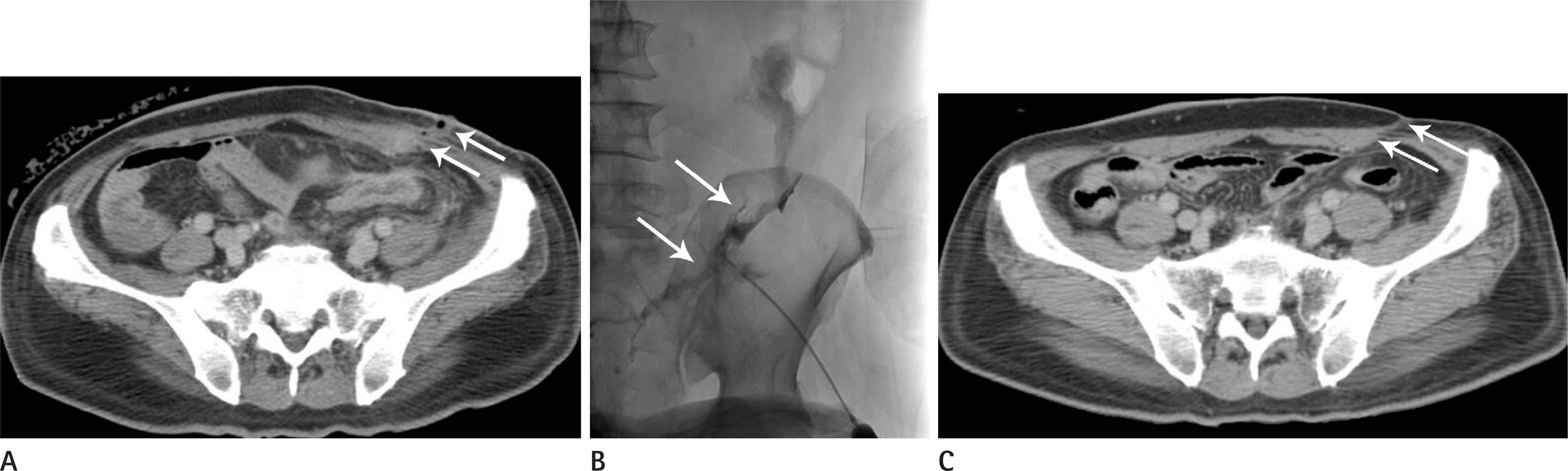 | Fig. 1.An external and low-output fistula after low anterior resection for rectal cancer. A. Axial CT image shows a colo-cutaneous fistula in the left lower abdomen (arrows). B. Fistulogram using iodinated water soluble contrast medium shows direct communication into the sigmoid colon (arrows). C. Two-month follow-up CT after external drainage shows disappearance of the fistulous tract (arrows). CT = computed tomography |
 | Fig. 2.An internal, ileo-colic fistula in Crohn's disease. A–C. Three dimensional reconstruction (A, B) and axial computed tomography (C) images after oral contrast ingestion show an ileo-colic fistula (arrows). D. Fistula tract between the ileum and the transverse colon is seen in a small bowel study (arrow). |
 | Fig. 3.External and high-output fistula (duodeno-cutaneous fistula) after surgical repair of traumatic duodenal perforation. A-C. In the initial (A) and follow-up contrast studies (B, C), an abscess cavity and a fistula tract (arrows) between the duodenum and skin are seen. D. Follow-up contrast study shows a completely closed fistula tract (arrow) after percutaneous radiologic drainage. |
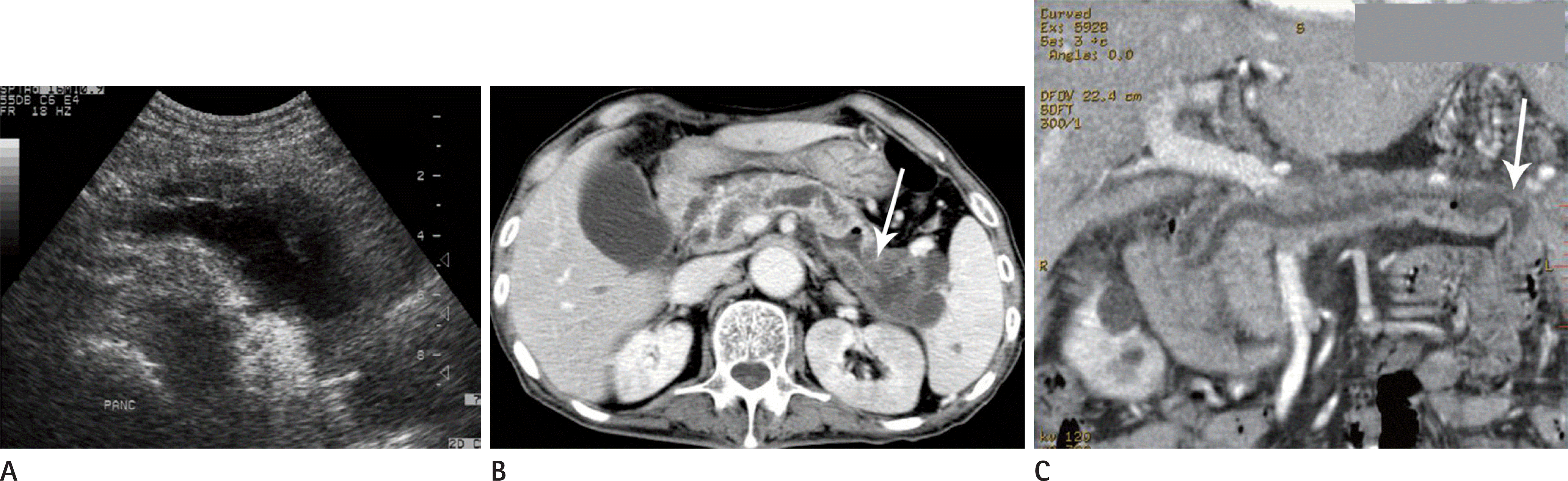 | Fig. 4.An internal, pancreatico-jejunal fistula in a malignant intramural papillary mucinous tumor. A. Initial, abdominal US image shows diffuse pancreatic duct dilatation. B, C. Axial (B) and three dimensional reconstruction CT (C) images taken three days after US show a pancreatico-jejunal fistula (arrows). Pancreatic duct dilatation is somewhat improved due to the fistula. CT = computed tomography, US = ultrasonography |
 | Fig. 5.External and high-output fistula after subtotal gastrectomy and gastroduodenostomy. A. Axial computed tomography image after the operation shows an air-containing abscess (arrows) around the operation site. B. After percutaneous drainage of the abscess, a fistula tract to the anastomotic site is noted (arrow). C. Upper gastrointestinal study performed 2 weeks after percutaneous drainage shows disappearance of the fistula tract. |
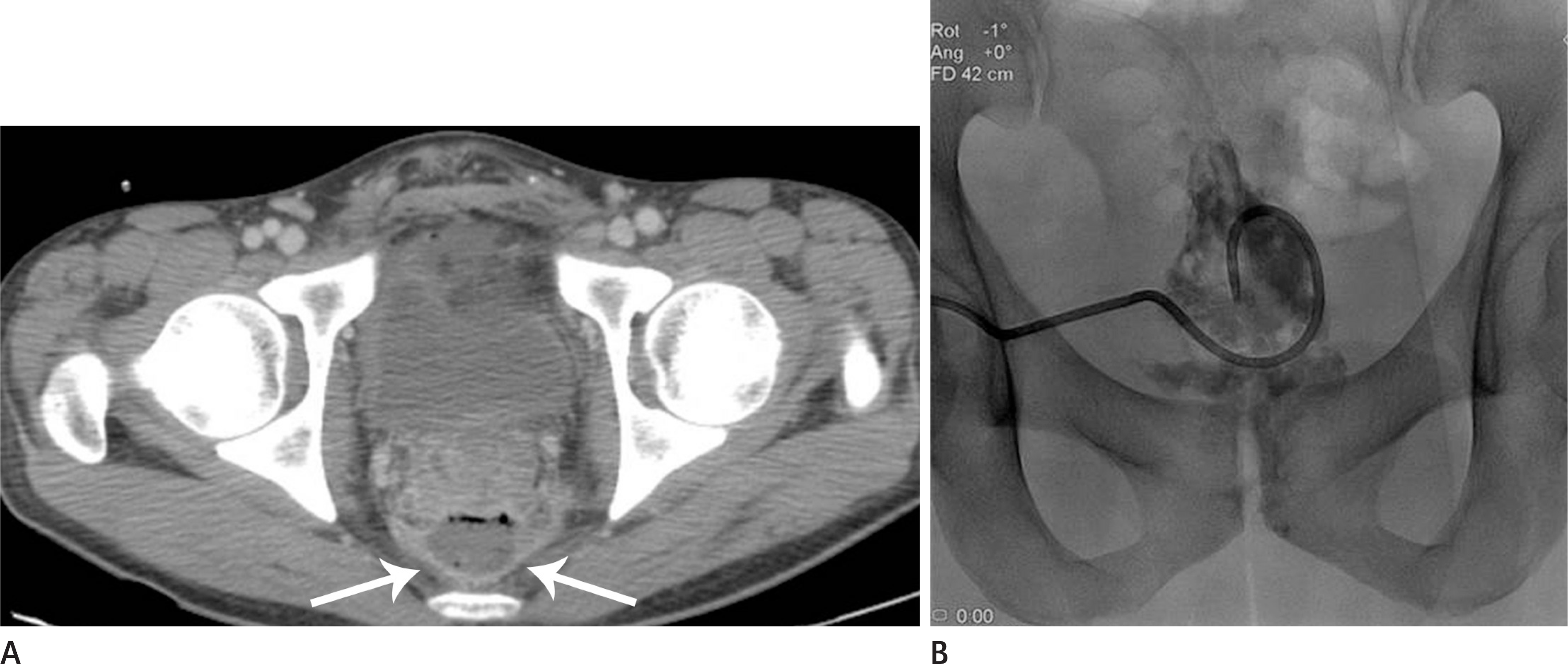 | Fig. 6.Transgluteal approach in the prone position for a presacral, pelvic abscess after proctocolectomy for rectal cancer. A. Axial computed tomography image shows complicated fluid collection in the presacral space (arrows). B. Percutaneous catheter drainage was performed via the left transgluteal route in the prone position. |
 | Fig. 7.Transvaginal approach for a deep-seated pelvic abscess after hysterectomy. A, B. Axial computed tomography image shows an irregular-shaped abscess cavity in the pelvic cavity and a fistula tract between the sigmoid colon and the abscess cavity (arrow). C. Transvaginal catheter drainage was performed under fluoroscopic guidance. |
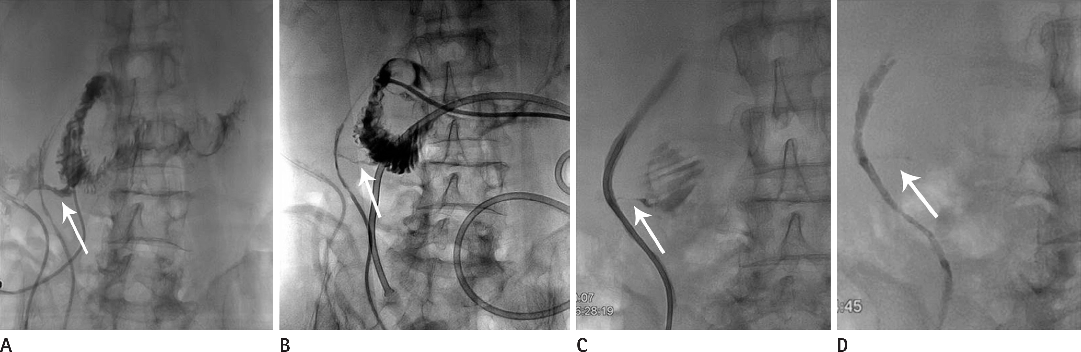
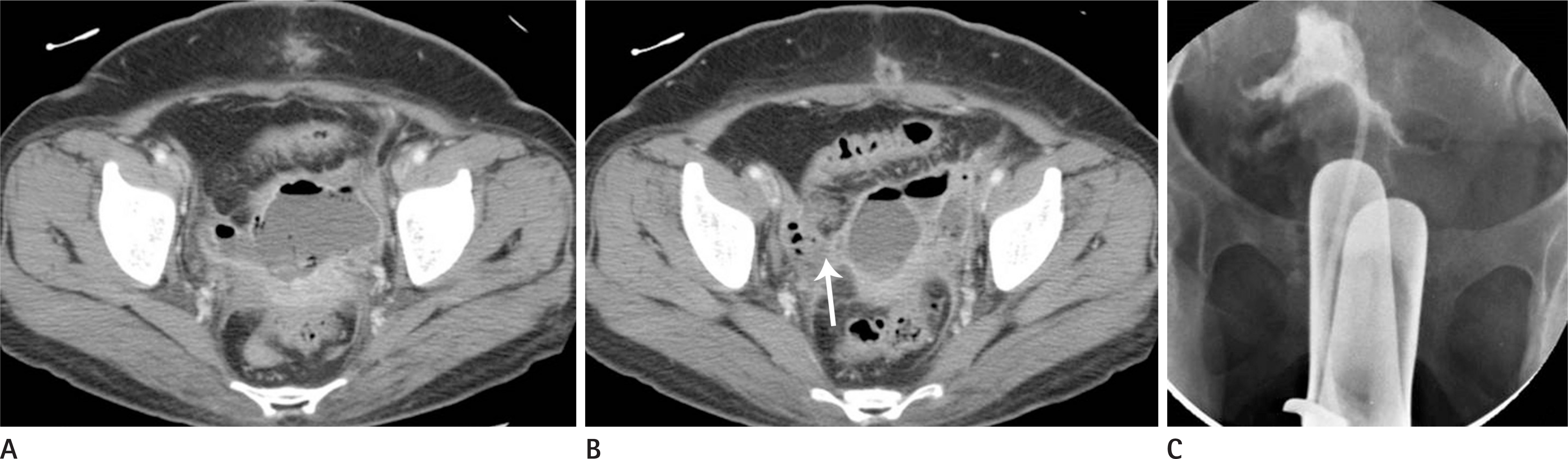
 | Fig. 8.Complex external and internal fistulae in a patient with necrotizing pancreatitis. A. Axial computed tomography image shows necrotizing pancreatitis and irregular fluid collection around the peripancreatic space (arrows). B. Contrast study via a drainage catheter shows complex fistulae among the abscess cavity, stomach, duodenum, and ascending colon. C. Upper gastrointestinal study after radiologic feeding jejunal tube insertion. The fistulae were closed 8 weeks after drainage tube and feeding jejunal tube insertion. |
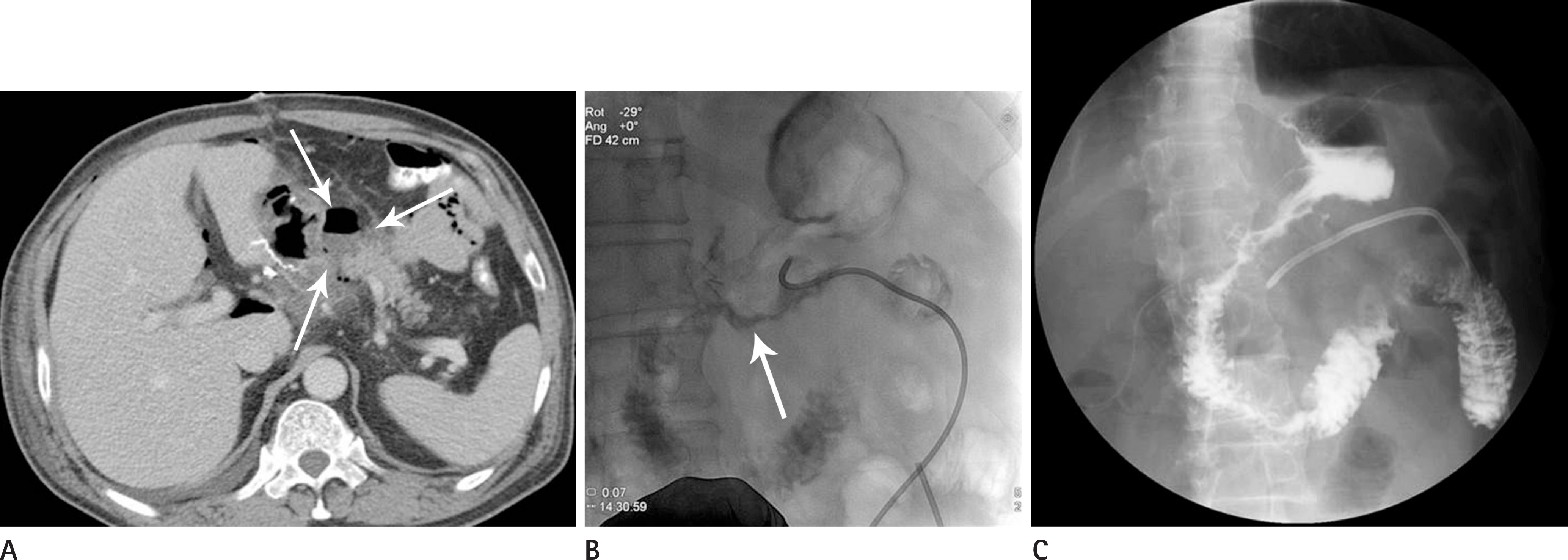
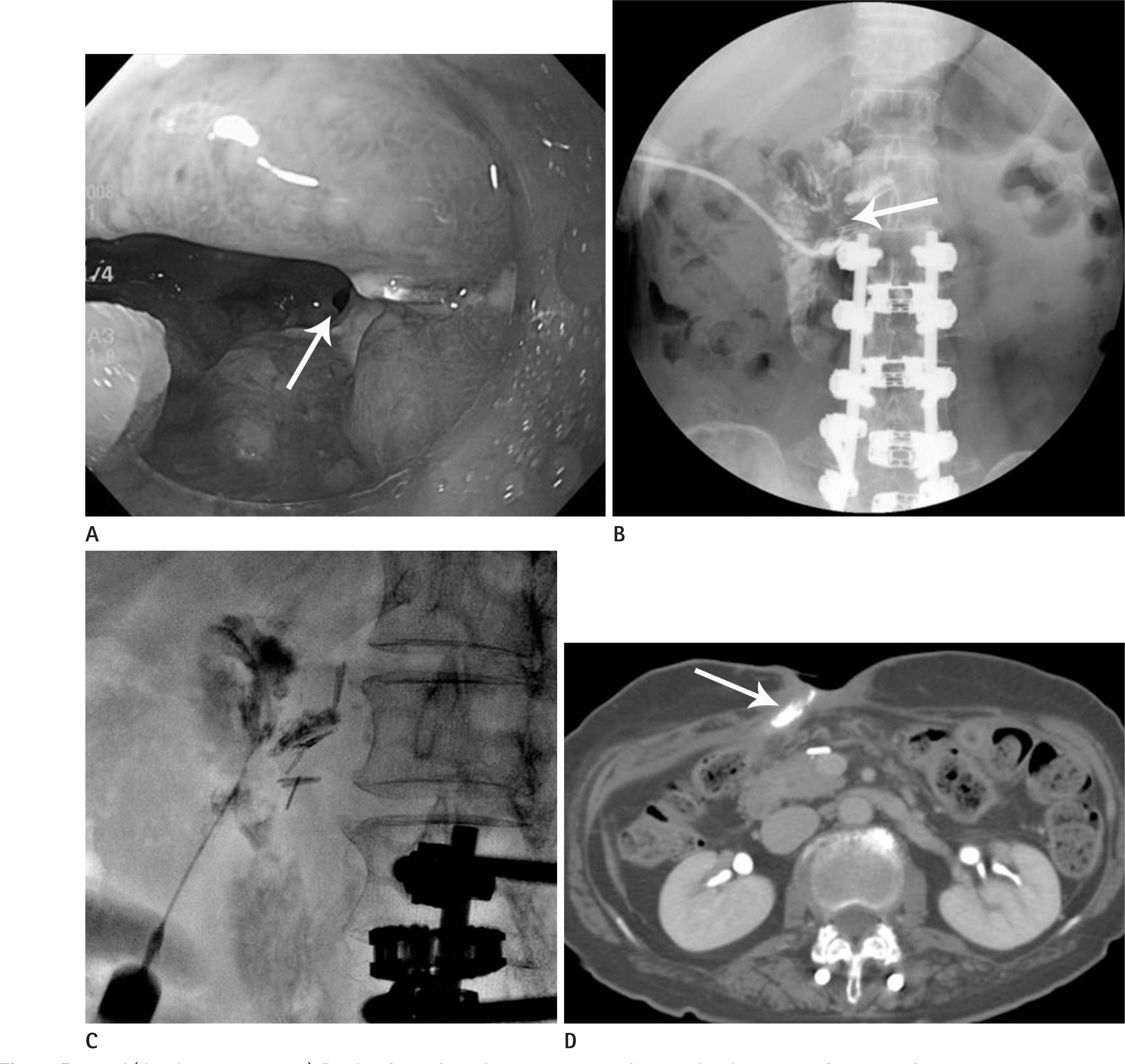 | Fig. 9.External (duodeno-cutaneous) fistula after subtotal gastrectomy and gastroduodenostomy for stomach cancer. A. Endoscopic image shows an internal fistula opening in the second portion of the duodenum (arrow). B. Sinogram of the fistula demonstrates a fistula tract to the second portion of the duodenum (arrow). C. Histoacryl glue (N-butyl-2-cyanoacrylate)-Lipiodol mixture (1:1) injection was performed for blockage of the fistula tract 8 weeks after conservative management. D. Follow-up computed tomography image shows blockage of the fistula tract with glue (arrow). |
 | Fig. 10.Histoacryl glue (NBCA) injection for a long-standing periappendiceal abscess –ascending colon fistula. A. Contrast study through a drainage catheter shows a fistula tract between the abscess cavity and the ascending colon (arrow). B. Histoacryl glue (NBCA)-Lipiodol mixture (1:1) injection was performed for blockage of the fistula tract 6 weeks after conservative management (arrow). C, D. Follow-up axial (C) and coronal (D) computed tomography images shows blockage of the previous fistula tract (arrows). NBCA = N-butyl-2-cyanoacrylate |
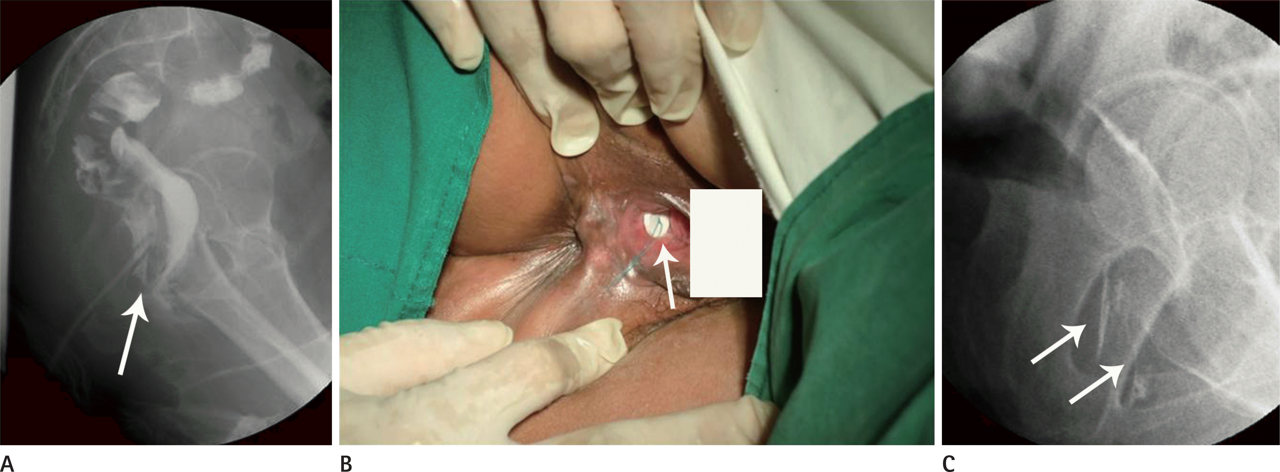 | Fig. 11.Histoacryl glue (NBCA) injection and rubber patch application for a longstanding recto-vaginal fistula due to radiation therapy. A. Colon study shows a recto-vaginal fistula (arrow). B, C. Rubber patch application (B) and histoacryl glue (NBCA)-Lipiodol mixture (1:1) injection (C) was performed (arrows). After the procedure, fecal discharge from the vagina was markedly decreased. NBCA = N-butyl-2-cyanoacrylate |
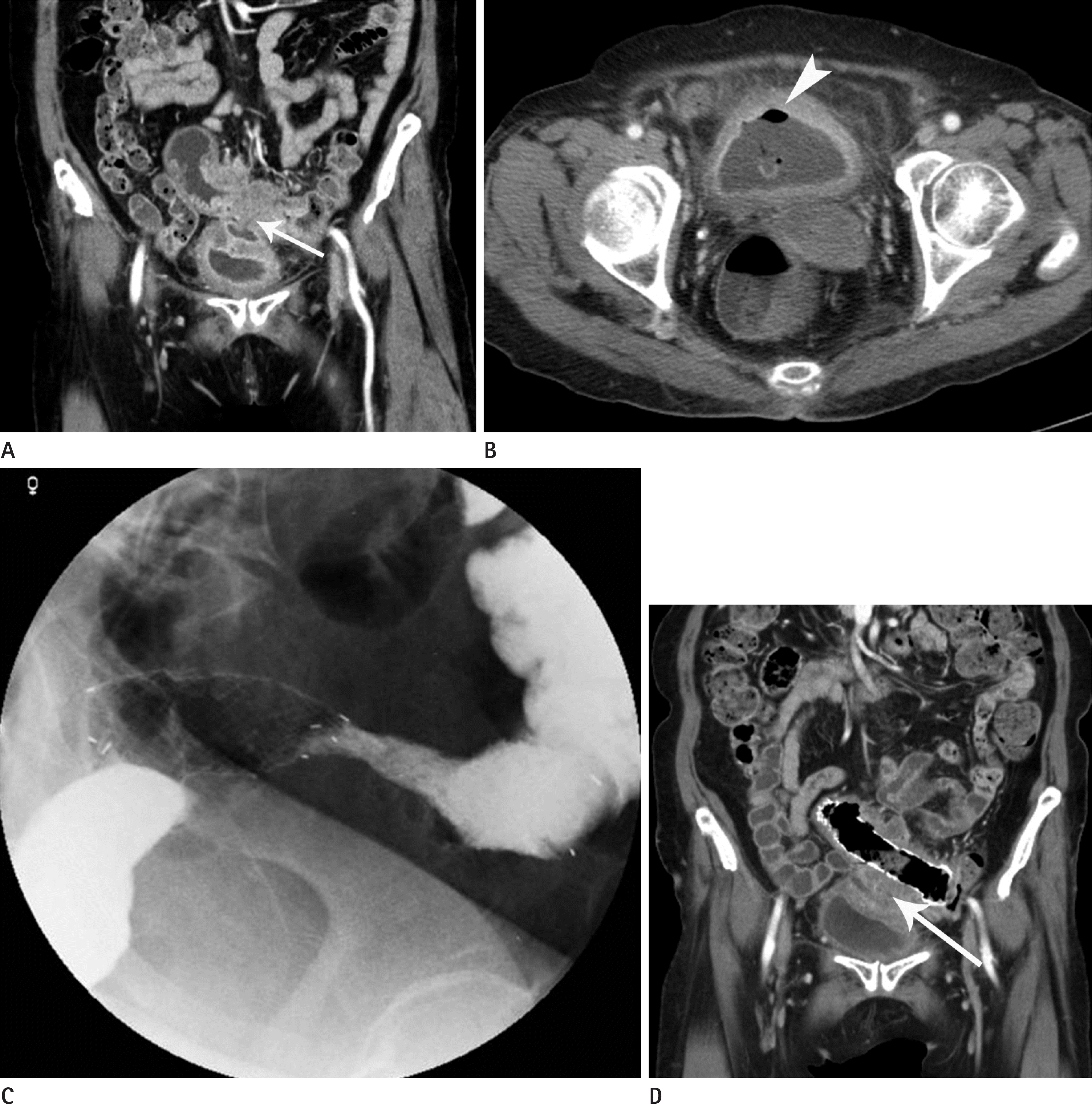 | Fig. 12.Covered metal stent placement for blockage of a sigmoid colo-vesical fistula in a patient with sigmoid colon cancer. A. Abdomen CT images show sigmoid colon cancer with a fistula tract between the sigmoid colon and the urinary bladder (arrow). B. Free air in the urinary bladder is seen due to the fistula (arrowhead). C. Covered metal stent placement (Hanaro, MI Tech, Seoul, Korea; 22 mm in diameter and 120 mm in length) was performed and the fistula tract was closed successfully. D. Follow-up coronal CT image 2 months after stent placement shows disappearance of the previous fistula tract (arrow). CT = computed tomography |
Table 1.
Classification of Gastrointestinal Fistulae
Table 2.
Etiologic Classification of Gastrointestinal Fistulae
Table 3.
Factors Affecting Spontaneous Closure of Gastrointestinal Fistulae
 XML Download