Abstract
Atypical teratoid/rhabdoid tumor (AT/RT) of spine usually reported to develop in the brain, while it rarely manifest in the spine. It consists of rhabdoid cells and is highly malignant. AT/RT appears at various sites throughout the body, such as in the central nervous system, liver, kidneys, abdomen, and soft tissues. Among them, spinal AT/RT is rare, and AT/RT of lumbar spine is extremely rare; only a few cases have been reported. We present the case of an AT/RT of lumbar spine in a 16-month-old boy.
Atypical teratoid/rhabdoid tumor (AT/RT) is uncommon malignant neoplasm that is usually reported in children and infants. It appears at numerous sites throughout the body, such as in the central nervous system (CNS), liver, kidneys, abdomen, and soft tissues. In the CNS, the posterior cranial fossa is the most common site for AT/RT. AT/RT of the spine is extremely rare, and fewer than five cases of spinal AT/RT in the lumbar region have been reported to our knowledge (1234). We present the case of an AT/RT of the lumbar spine in a 16-month-old boy.
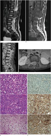
A 16-month-old boy without an underlying medical history presented with weakness in both legs since two weeks. He was unable to walk or sit down plump since two days. He had experienced a severe cold, three weeks before visiting the hospital. Physical examination revealed muscle tone loss in the lower extremities and no increased deep tendon reflex. Magnetic resonance imaging (MRI) of the lumbosacral spine showed a dumbbell-shaped mass in the spinal canal on the right at the level of L1–L3, extending to the right lumbar plexus through the right intervertebral foramen at the level of L2–L3. The mass was heterogeneous with iso - hyper signal intensity on T2-weighted images. There was heterogeneous enhancement on contrastenhanced images (Fig. 1A-D). Imaging findings of brain MRI were normal. On the day that MRI was conducted, surgical resection of the tumor and L1–L4 laminoplasty were conducted. In addition to laminoplasty, retroperitoneal exploration was conducted to rule out mass invasion. No metastasis was detected in the retroperitoneal space or kidneys.
Histologically, the tumor was composed of rhabdoid cells, that comprised eosinophilic cytoplasm with eccentric nuclei. Some cells comprised abundant eosinophilic cytoplasm and prominent nucleoli. Cords of tumor cells appeared in a myxoid matrix, resembling chondroid differentiation (Fig. 1E).
Immunohistochemical staining for integrase interactor 1 (INI1), the product of the SMARCB1 gene, showed loss of expression in the nuclei of tumor cells. Immunohistochemical staining for epithelial membrane antigen (EMA) and cytokeratin were positive (Fig. 1F).
These morphological and immunohistochemical findings were the basis for the diagnosis of an AT/RT.
The patient was scheduled for radiation therapy.
AT/RT is highly malignant neoplasm that originate in the CNS. It usually occurs in the brain and spinal AT/RT is rare. Originally, AT/RT was called malignant rhabdoid tumor. Beckwith and Palmer (5) first described it as an aggressive variant of Wilms' tumor arising from the kidneys. It was defined as distinguished CNS tumor and given the name “atypical teratoid/ rhabdoid tumor” in 1996 (6). Among spinal AT/RT, lumbar region localization is extremely rare and fewer than five cases have been reported (1234).
A review of the four documented cases of lumbar spinal AT/RTs revealed an age range of 16 months to seven years. Three of them were in the intradural extramedullary region, and in the remaining case in an 18-month-old infant, the AT/RT was in the paraspinal region with intraspinal extension, like our case. Three cases were in boys and one was in a girl.
AT/RT is usually reported as heterogeneous intense mass on T2-weighted images with internal hyperintense foci on T1- weighted images. The tumor is located in intramedullary or intradural extramedullary region in most cases (7).
Histopathologically, AT/RT is composed of markedly proliferating rhabdoid cells, that exhibit a high nuclear-to-cytoplasmic ratio, and malignant epithelial and mesenchymal components that lack divergent tissue differentiation characteristics. On immunostaining, AT/RT reveals immunoreactivitiy to EMA, vimentin, and smooth muscle antigen, while showing negative immunoreactivity to INI1. Chromosome 22q11 abnormalities are of crucial diagnostic value (8).
Differential diagnoses depend on the origin and location of the lesion. For lesions of the extramedullary region, malignant nerve sheath tumors, and myxopapillary ependymomas should also be considered. For lesions of the intramedullary region, ependymomas, gangliogliomas, and astrocytomas should be considered. Whole-CNS imaging should be considered because the lesion may also be metastatic.
In case of AT/RT, surgery and post-operative chemotherapy are considered. For patients with metastatic disease, high-dose chemotherapy may be useful. Additional radiation therapy may be administered (910).
AT/RT is malignant and results in poor patient prognosis. Published reports on CNS AT/RTs in those younger than age three indicate that the overall two-year survival rate is less than 15% (89). However, there are only limited data from spinal AT/RT due to its rarity.
In this report, we presented a case of an AT/RT of the lumbar spine, presenting as a dumbbell-shaped mass in the spinal canal extending to the right lumbar plexus. Considering its diagnostic difficulty due to non-specific imaging findings and its rarity, early diagnosis of the AT/RT and aggressive treatment should be conducted as soon as possible. Long-term and strict followups are recommended.
Figures and Tables
 | Fig. 1Atypical teratoid/rhabdoid tumor of lumbar spine in an infant.
A. T2-weighted sagittal image shows a heterogeneously isointense and hyperintense mass in the spinal canal at the level of L1-L3.
B. The mass exhibits isointensity on the T1-weighted sagittal image.
C. The gadolinium-enhanced, T1-weighted, fat-suppressed sagittal image shows heterogeneous enhancement of the mass.
D. The gadolinium-enhanced, T1-weighted, fat-suppressed axial image shows a dumbbell-shaped mass in the spinal canal on the right, extending to the right lumbar plexus through the right intervertebral foramen.
E. The tumor shows a variety of histologic patterns. The tumor cells have eosinophilic cytoplasm with eccentric nuclei; these are typical rhabdoid features. Some areas show large tumor cells with abundant eosinophilic cytoplasm and prominent nucleoli. Cords of tumor cells appear in a myxoid matrix resembling chondroid differentiation (hematoxylin and eosin stain, a: × 200, b: × 200, c: × 100).
F. Immunohistochemical staining for integrase interactor 1, the product of the SMARCB1 gene, shows loss of expression in the nuclei of tumor cells, compared to retained staining of the nuclei of inflammatory cells. The tumor cells are positive for epithelial membrane antigen and cytokeratin (a: integrase interactor 1 stain, × 200, b: epithelial membrane antigen stain, × 200, c: cytokeratin stain, × 200).
|
References
1. Yang CS, Jan YJ, Wang J, Shen CC, Chen CC, Chen M. Spinal atypical teratoid/rhabdoid tumor in a 7-year-old boy. Neuropathology. 2007; 27:139–144.

2. Agrawal A, Bhake A, Cincu R. Giant lumbar paraspinal atypical teratoid/rhabdoid tumor in a child. J Cancer Res Ther. 2009; 5:318–320.

3. Dhir A, Tekautz T, Recinos V, Murphy E, Prayson RA, Ruggieri P, et al. Lumbar spinal atypical teratoid rhabdoid tumor. J Clin Neurosci. 2015; 22:1988–1989.

4. Chao MF, Su YF, Jaw TS, Chiou SS, Lin CH. Atypical teratoid/ rhabdoid tumor of lumbar spine in a toddler child. Spinal Cord Ser Cases. 2017; 3:16026.

5. Beckwith JB, Palmer NF. Histopathology and prognosis of Wilms tumors: results from the first national Wilms' tumor study. Cancer. 1978; 41:1937–1948.
6. Rorke LB, Packer RJ, Biegel JA. Central nervous system atypical teratoid/rhabdoid tumors of infancy and childhood: definition of an entity. J Neurosurg. 1996; 85:56–65.

7. Moeller KK, Coventry S, Jernigan S, Moriarty TM. Atypical teratoid/rhabdoid tumor of the spine. AJNR Am J Neuroradiol. 2007; 28:593–595.
8. Biegel JA, Tan L, Zhang F, Wainwright L, Russo P, Rorke LB. Alterations of the hSNF5/INI1 gene in central nervous system atypical teratoid/rhabdoid tumors and renal and extrarenal rhabdoid tumors. Clin Cancer Res. 2002; 8:3461–3467.


 PDF
PDF ePub
ePub Citation
 Print
Print


 XML Download