

 PDF
PDF ePub
ePub Citation
 Print
Print


Abstract
Although bimaxillary surgery enhances patients' self-esteem and mood by improving their facial appearance, surgical outcome assessments for this procedure are limited. This preliminary study investigated differences in brain activity on functional magnetic resonance imaging (fMRI) during self-face evaluation before and after bimaxillary surgery. Three patients (1 man and 2 women, age range: 20–27 years) underwent fMRI while viewing self-face images before and after bimaxillary surgery for maxillofacial deformity. The activation in the left postcentral gyrus, and medial orbital frontal cortex was significantly great in response to after-surgery self-face images compared to before-surgery images. Our preliminary results may facilitate the development of an objective measure for patient satisfaction after orthognathic surgery including bimaxillary surgery.
Go to : 
References
1. Hwang K, Choi YB. Postoperative monitoring following jaw surgery is essential. Arch Plast Surg. 2013; 40:66–67.

2. Khechoyan DY. Orthognathic surgery: general considerations. Semin Plast Surg. 2013; 27:133–136.
3. Murphy C, Kearns G, Sleeman D, Cronin M, Allen PF. The clinical relevance of orthognathic surgery on quality of life. Int J Oral Maxillofac Surg. 2011; 40:926–930.
4. Ueno A, Ito A, Kawasaki I, Kawachi Y, Yoshida K, Murakami Y, et al. Neural activity associated with enhanced facial attractiveness by cosmetics use. Neurosci Lett. 2014; 566:142–146.
5. O'Doherty J, Winston J, Critchley H, Perrett D, Burt DM, Dolan RJ. Beauty in a smile: the role of medial orbitofrontal cortex in facial attractiveness. Neuropsychologia. 2003; 41:147–155.
6. Aharon I, Etcoff N, Ariely D, Chabris CF, O'Connor E, Breiter HC. Beautiful faces have variable reward value: fMRI and behavioral evidence. Neuron. 2001; 32:537–551.
7. Stalnaker TA, Cooch NK, Schoenbaum G. What the orbitofrontal cortex does not do. Nat Neurosci. 2015; 18:620–627.
8. Platek SM, Loughead JW, Gur RC, Busch S, Ruparel K, Phend N, et al. Neural substrates for functionally discriminating self-face from personally familiar faces. Hum Brain Mapp. 2006; 27:91–98.
9. Shen H, Chau DK, Su J, Zeng LL, Jiang W, He J, et al. Brain responses to facial attractiveness induced by facial proportions: evidence from an fMRI study. Sci Rep. 2016; 6:35905.
10. Martín-Loeches M, Hernández-Tamames JA, Martín A, Urrutia M. Beauty and ugliness in the bodies and faces of others: an fMRI study of person esthetic judgement. Neuroscience. 2014; 277:486–497.
Go to :
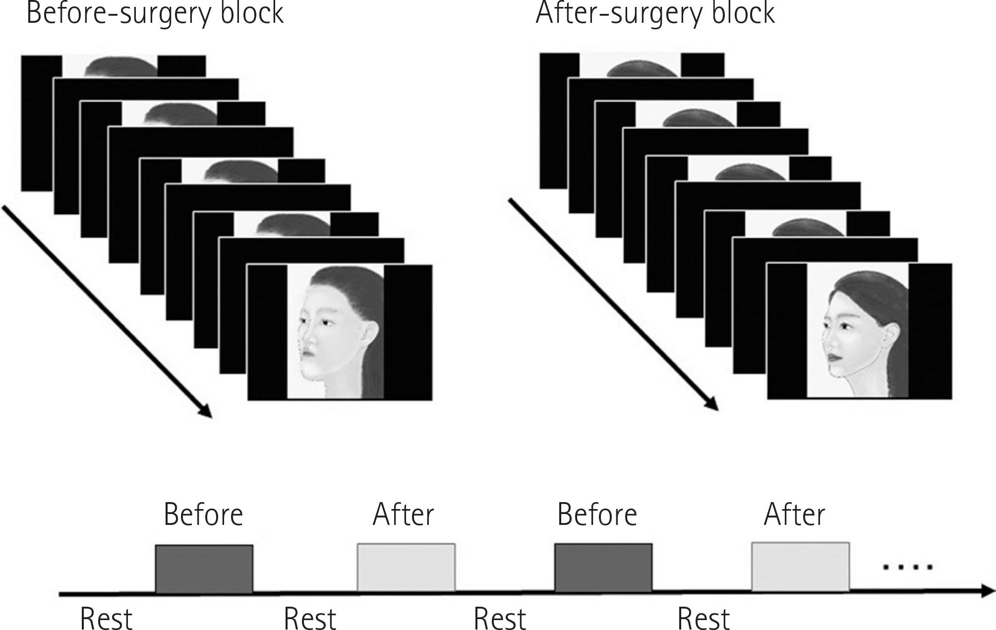 | Fig. 1.Functional magnetic resonance imaging experimental design. Before surgery blocks and after-surgery blocks were alternated 4 times in a random order in each session. Each block consisted of 10 images presented for 1 s each. Once the image had disappeared, a small fixation cross appeared in the center of the screen. The time required for each block was 30 s, and a 30 s rest block was inserted between the activation blocks (before surgery blocks and after surgery blocks). During the rest block, a small fixation cross on a black background was presented. |
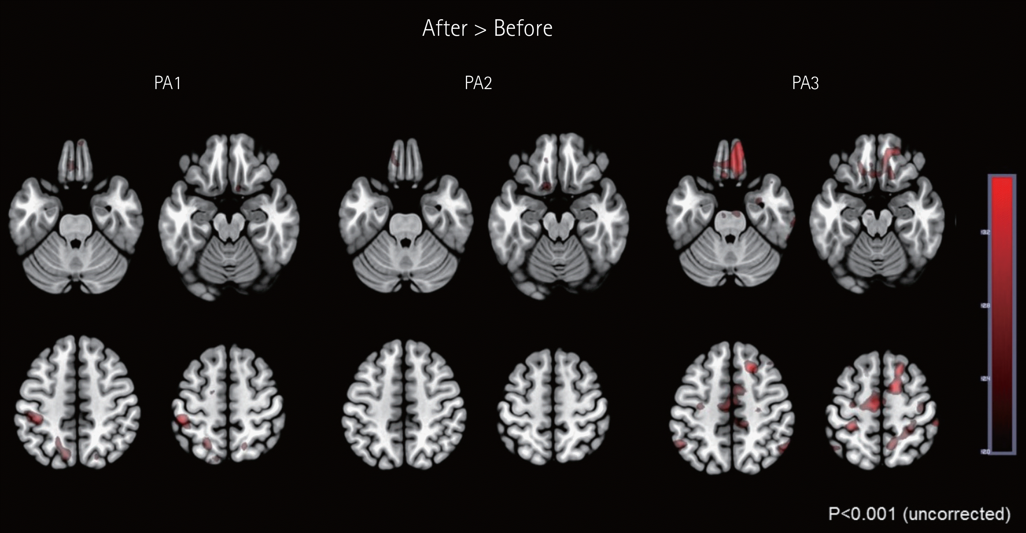 | Fig. 2.Increased activation in response to after-surgery faces versus before-surgery faces. Images of faces taken after surgery revealed greater activation in the left postcentral gyrus, cerebellar vermis, and right medial orbitofrontal cortex than images taken before surgery (p < 0.001 and a cluster size of 10 voxels) and left medial orbitofrontal cortex (p < 0.01 and a cluster size of 5 voxels). Brain left is on the figure left. PA = patient |
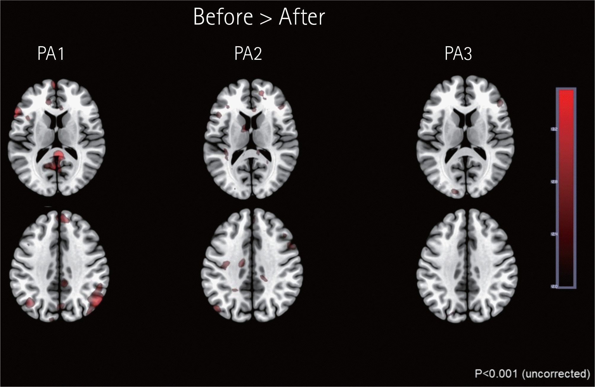 | Fig. 3.Increased activation in response to before-surgery faces versus after-surgery faces. Images of faces taken before surgery revealed greater activation in the right posterior cingulate cortex, bilateral inferior parietal lobules, left superior frontal gyrus, bilateral medial frontal gyri, left inferior frontal gyrus, right parahippocampal gyrus, left middle frontal gyrus, and left middle temporal gyrus than those taken after surgery (p < 0.001 and a cluster size of 10 voxels). Brain left is on the figure left. PA = patient |
Table 1.
Montreal Neurological Institute Coordinates of Neural Activation in 3 Subjects
| After > Before | Before > After | |||
|---|---|---|---|---|
| Region | X, Y, Z | Region | X, Y, Z | |
| Case 1 | Left postcentral | –46, −34, 54 | Right posterior cingulate | 4, −40, 16 |
| Left Precuneus | –10, −42, 74 | Right angular gyrus | 46, −68, 46 | |
| Right medial orbital frontal (BA11) | 10, 14, −18∗ | Right superior medial frontal gyrus | 4, 52, 42 | |
| Left superior medial frontal gyrus | –2, 62, 10 | |||
| Left supplementary motor area | –2, 14, 66 | |||
| Left inferior frontal gyrus (triangular) | –40, 10, 24 | |||
| Left angular gyrus | –40, 68, 40 | |||
| Case 2 | Vermis 4–5 | 0, −50, 6 | Calcarine, right | 24, −48, 4 |
| Left medial orbital∗ frontal (BA11) | –12, 14, 22∗ | Middle frontal, left | –34, 46, 2 | |
| Case 3 | Right medial orbital frontal (BA11) | 10, 50, −26 | Left medial frontal gyrus | –4, 54, −12 |
| 20, 50, −16∗ | Left middle temporal gyrus | –48, −60, −2 | ||
Peak voxel sizes for all regions were obtained from a whole-brain random-effects contrast of before surgery and after surgery face stimuli. p < 0.001, uncorrected at the voxel level with extent of 10 contiguous voxels or more. ∗p < 0.01, uncorrected at the voxel level with extent of 5 contiguous voxels or more. BA = brodmann area
 XML Download