

 PDF
PDF ePub
ePub Citation
 Print
Print


INTRODUCTION
Several imaging modalities can be used for the detection of inflammation, such as computed tomography (CT), magnetic resonance imaging (MRI), gamma scintigraphy and positron emission tomography/computed tomography (PET/CT) (1). Among the nuclear medicine imaging modalities, three-phase bone scan, radioisotope-labeled white blood cell (WBC) scan, and 18F-fluorodeoxyglucose PET/CT are commonly used to detect the inflammation site (12). The 18F-sodium-fluoride (18F-NaF) bone PET/CT is preferred as it provides additional early image acquisition within the first 10 min after injection and therefore, can be used to detect osteomyelitis (3). These earlyphase scan images can substitute for the perfusion and blood pool phases of the three-phase bone scan (3). Moreover, conventional imaging modalities including CT and MRI have a limited role in evaluating patients with metallic implants. In contrast, PET/CT can be used to overcome this artifact using non-attenuation corrected (NAC) images (4).
We report a case of a patient with increased serum C-reactive protein (CRP) levels without a definite inflammatory focus after spinal operation. Using two-phase 18F-NaF bone PET/CT including early-phase scan and additional NAC images, we accurately detected the inflammation site and evaluated early treatment response.
CASE REPORT
A 76-year-old woman presented with low back pain for 3 years. She was diagnosed with spinal stenosis at the L3–S1 spinal level. She underwent partial discectomy of the L3–S1 spine and posterior lumbar interbody fusion of the L3–L5 spine. An additional operation to remove postoperative hematoma was performed six days after surgery. After hematoma removal, the blood tests revealed an elevated CRP level of 60.82 mg/L (normal range; 0–3.0 mg/L). Elevation of the CRP level persisted for more than 2 weeks. Chest X-ray and urine examination were performed to evaluate the inflammatory focus, however, no abnormal findings were found. Wound cultures obtained from the operation site following irrigation reported no bacterial growth.
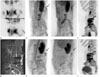
99mTc-labeled WBC scan and two-phase 18F-NaF bone PET/CT were performed to assess the inflammatory focus. The WBC scan revealed radiotracer accumulation in the perispinal space from the kidney to the sacral level, suggesting active inflammation (Fig. 1A). The next day, a two-phase 18F-NaF bone PET/CT with early-phase scan was performed to exclude the possibility of bone involvement of soft tissue inflammation. Immediately after intravenous injection of 185 MBq of 18F-NaF, using Biograph mCT 128 scanner (Siemens Healthcare, Knoxville, TN, USA), early phase images were acquired with static acquisition (2 min per bed position). Early phase scan revealed an increase in soft tissue uptake around the hematoma removal site and the spinal operation site without involving the spine. NAC PET/CT images also showed an increase in radiotracer uptake in the same area (Fig. 1B), indicating that the activity was not due to a metal artifact. Forty-five minutes after intravenous administration of 18F-NaF, a standard 18F-NaF PET/CT was performed. The standard 18F-NaF PET/CT images indicated an increase in radiotracer uptake at the operation site of spine (Fig. 1C). Diffuse radiotracer uptake was observed in the vertebral body and the epiphyseal plate, suggesting a post-operative change. Subsequently, she underwent L-spine MRI via the Dixon technique, which showed post-operative hematoma at the interface between the back muscle and the subcutaneous fat layer, in addition to low signal intensity due to metal artifact (Fig. 1D).
Following a diagnosis of soft tissue inflammation around the operation site, intravenous rifampin treatment was started. Two months after the antibiotic treatment, the serum CRP level decreased to the normal range (2.23 mg/L) and follow-up 18F-NaF bone PET/CT was performed. Early-phase scan images demonstrated a decrease in the intensity of uptake of pre-existing radiotracer, however, a mild uptake was still observed (Fig. 1E, left). In contrast, markedly decreased radiotracer uptake without discernible residual uptake was seen on NAC PET/CT images (Fig. 1E, right). The standard 18F-NaF PET/CT images (Fig. 1F) revealed a diffused radiotracer uptake due to post-operative changes without any significant difference from previous PET/CT findings.
DISCUSSION
The utility of 18F-NaF bone PET/CT has been established in several studies that evaluated osteomyelitis as well as bone metastasis (5). The intravenously administered 18F-NaF is adsorbed into the hydroxyapatite crystalline structure, which is comprised of calcium and phosphate crystals (6). According to Freesmeyer et al. (7), early-phase images of 18F-NaF PET/CT show blood distribution similar to the blood-pool phase in the three-phase bone scan.
In this case, 18F-NaF bone PET/CT elucidated the inflammation in the WBC scan and excluded the possibility of osteomyelitis. Moreover, 18F-NaF bone PET/CT images show higher spatial resolution and facilitate the evaluation of the spine region, compared with WBC scan (38). Until now, MRI has been the preferred modality for evaluation of soft tissue lesions (9). However, in many cases treated with orthopedic surgery, metals are used, which cause artifacts and limit the accuracy of MRI (10). In our case, the Dixon technique was used to overcome artifacts, however, MRI images still showed a limitation in evaluation of the inflammation site. During PET/CT, the CT-based attenuation correction using the Hounsfield unit number leads to overestimation of radiotracer uptake in patients carrying metallic implants (4). Comparison with NAC PET images allows the interpretation of metal-induced artifacts (4).
In this case, initial early-phase 18F-NaF bone PET/CT images showed radiotracer uptake at the operation site. However, it should be confirmed whether the radiotracer uptake detected in attenuation-corrected PET/CT images indicates a true lesion because the patient underwent surgery with metallic implants in place. NAC images showed a pattern similar to the attenuation-corrected images, confirming the finding of a true inflammatory lesion. The diagnosis of soft tissue inflammation around the operation site without bone involvement was established using a two-phase 18F-NaF bone PET/CT. Follow-up with attenuation-corrected imaging of early-phase 18F-NaF bone PET/CT showed a decrease in uptake intensity of pre-existing radiotracer uptake, although a mild radiotracer uptake was still found. However, the NAC images showed a markedly decreased radiotracer uptake without a residual uptake in the lesion, suggesting resolution of active inflammation. The standard 18F-NaF PET/CT images showed diffusely increased radiotracer uptake around the operation site without any interval change after antibiotic treatment, suggesting the limited role of standard bone PET/CT in evaluating bony lesions postoperatively.
In the present case, 18F-NaF bone PET/CT images with early phase scan were used to elucidate the inflammation site. The strategy facilitated accurate diagnosis and treatment response evaluation using NAC images without the effect of metal artifacts.
 XML Download