

 PDF
PDF ePub
ePub Citation
 Print
Print


INTRODUCTION
Extrapleural solitary fibrous tumor, formerly referred to as “hemangiopericytoma” is a rare soft tissue vascular tumor arising from the capillary pericytes which has elevated makignancy potential. The tumor can be found throughout the entire body that contain capillaries. Recently solitary fibrous tumor, hemangiopericytoma, lipomatous hemangiopericytoma and giant cell angiofibroma have been categorized as extrapleural solitary fibrous tumors (12). In prior reports, extrapleural solitary fibrous tumors, originating from the spleen, are described as extremely rare entities and only 19 cases have been reported (3). In this report, we describe a rare case of a patient with an splenic extrapleural solitary fibrous tumor. We also included a review of the literature, concentrating on radiologic imaging findings of extrapleural solitary fibrous tumors arising in the spleen.
CASE REPORT
A 35-year-old woman with nausea and malaise was admitted to the gastroenterology and hepatology outpatient department of the hospital. Her aspartate transaminase and alanine transaminase levels were elevated to 271 and 940 IU/L, respectively. Her checked total bilirubin level was also elevated to 4.96 mg/dL on initial laboratory examination. She had no significant past medical or family history and her physical examination results were not notable. She was treated under the diagnosis of hepatitis A.
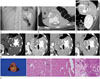
The patient underwent conventional radiography and computed tomography (CT) of the abdomen. On conventional radiography of the abdomen, a multilobulated, irregular calcified lesion with internal multifocal radiolucent foci was incidentally found in the left upper quadrant of her abdomen (Fig. 1A).
In the abdomen CT scan, a localized multilobulated mass was found in the spleen. The mass was measured to be approximately 7.8 × 5.6 × 5.8 cm in size. Large irregular-shaped dense calcifications with nothing in common, including size, shape, and margin were scattered throughout the entire mass on the pre-contrast enhancement. The biggest calcification which showed eccentric shape was located in the central portion. Some calcifications were located peripherally and others surrounded the intratumoral solid portion. The solid portion, which was accompanied by a calcified lesion in the mass, showed approximately 40 Hounsfield unit (HU) on the pre-contrast CT, 95 HU in the arterial phase of the post-contrast CT and approximately 140 HU in the venous and delayed phase. The venous and delayed phase displayed a wider enhancing area than did the arterial phase. In addition, a non-enhanced a low-attenuation lesion (approximately −80 HU to −20 HU) suggesting a fatty component was located in the central portion of this mass. The mass was confined to the spleen without invasionof adjacent organs and displayed no feeding artery or drainage vein except splenic vessels. There were no enlarged or enhanced lymph nodes in the abdomen found through the CT scan (Fig. 1B–D).
A splenic tumor, such as a teratoma or an angiosarcoma, was suspected on the first inspection because fat attenuation and irregular calcification in the mass was seen in the CT scan. We recommended further workup such as ultrasound and MRI. However, laparoscopic splenectomy, including the mass, was performed without any other radiologic examinations. The surgery was successfully completed without complication.
Pathologically, the mass proved to be an extrapleural solitary fibrous tumor. The mass was noted to have a smooth, bulging and grayish-brown external capsule in the gross findings. In the section, the mass had internal bizarre-shaped calcification and relatively well-defined solid lesion with some hemorrhage, but with no fatty component. There was no direct invasion to adjacent organs which was suspected on CT images.
In immunohistochemical staining, the tumor cells showed partly positive reaction on CD34 and SMA. Tumor cells displayed a diffuse positive reaction on vimentin, and a negative reaction on EMA, S100P, and CK which was pathologically compatible with an extrapleural solitary fibrous tumor (Fig. 1E).
DISCUSSION
Extrapleural solitary fibrous tumor previously known as “hemangiopericytoma” is characterized as a rare vascular tumor with very high malignant potential. The tumor was first described by Stout and Murray in 1942 and has been known to evolve from Zimmerman pericytes associated with capillaries and venules. Recent histopathologic studies have suggested that it originates from alternative cells such as fibroblasts. Additionally, the new 2013 World Health Organization (WHO) classification of tumors of the soft tissue abandoned the term “hemangiopericytoma” and merged it to solitary fibrous tumor, lipomatous hemangiopericytoma and giant cell angiofibroma under the category of extrapleural solitary fibrous tumor (12).
Extrapleural solitary fibrous tumor commonly form in adults from 20 years to 70 years old, with a median age of people in their 40s. It has similar prevalence in men and women (4). This tumor is most common in the pelvic retroperitoneum (39%) and emerges in the following order: extremities (22%), dura mater (19%), head and neck (11%), and the thorax (8%) (3).
Extrapleural solitary fibrous tumors in the spleen, as a primary tumor, was first reported in 1989 by Guadalajara Jurado et al. (5) and so far only 19 patients have been reported in the literature (3) to have this tumor. Patients with splenic extrapleural solitary fibrous tumors have variable clinical presentations such as a pain, lump, rupture, hemorrhage, and abscess (3567).
Prior literature has described the general CT findings of splenic extrapleural solitary fibrous tumor as a large, well-defined and multilobulated mass that is highly vascularized (8). The presence of large collateral feeding vessels is a useful distinguishing feature of solitary fibrous tumors (9). If necrosis, calcifications which are frequent in large tumors, and invasion to adjacent structures are shown; this is suggestive of a malignant form (4). Even if these morphologic characteristics are present, it is difficult to consider this tumor on the first impression when it originates from the spleen because of it is rare. Our case of splenic extrapleural solitary fibrous tumor showed general features on the CT. Moreover multiple internal irregular-shaped calcifications and some fat components were inconsistent with the reported general features. The possibility that the tumor has a malignant potential on radiologic findings is well worth considering because calcification is rare in this type of tumor (10) and the distribution pattern of calcification is eccentric. However, a pathologist reported that it was not a malignant form. In this case, only a CT scan was performed. Obviously, CT is helpful for the evaluation of features of intratumoral calcification, such as size, shape, and margin. However, considering the rarity of the disease, there are limitations regarding accurate information and diagnosis of the lesion only through CT. Additional examinations, including sonography and MRI, can provide supplementary information about the tumor. Although radiologic multimodalities may be useful for the diagnosis, it cannot be confirmed by radiologic studies only.
It is difficult to predict the prognosis and clinical course of extrapleural solitary fibrous tumors. In prior literature, it has been suggested that large size (> 5 cm), numerous mitoses (> 4 mitoses/10 high-powered field), cellular atypia, presence of necrosis and/or hemorrhage can be useful for differentiating the malignant from the benign forms (4). Surgical resection, including the tumor, is recommended as the treatment of choice. Wignall et al. (9) reported the metastasis rate of extrathoracic solitary fibrous tumors is 11%. Thus, the early radiologic detection and the diagnosis of extrapleural solitary fibrous tumor originating from the spleen is important for setting the establishing treatment and achieving better prognosis. Radiologists need to consider extrapleural solitary fibrous tumors as a diagnosis when differentiating splenic masses that contain irregular-shaped dense calcification.
 XML Download