Abstract
Deep vein thrombosis (DVT), which usually occurs in the lower extremities, is the presence of a blood clot within a deep vein that causes symptoms by breaking the venous return. Many cases of calf vein DVT are asymptomatic, or display only mild symptoms. But in the case of a proximal DVT, it affects the venous flow through the entire lower extremity, which results in a post-thrombotic syndrome, or a pulmonary embolism, if the proper treatment isn't performed. The diagnosis of the DVT is made by a radiologic examination. An ultrasound is often used as a first line of diagnosis, but on the other hand, computed tomography venography has also been gaining traction as an alternative method. If diagnosed, finding the cause of the DVT is important, and in the case of a symptomatic proximal DVT, the combination of anticoagulation and interventional treatment can be used towards the recovery of the venous return, preventing complications.
References
1. Black CM. Anatomy and physiology of the lower-extremity deep and superficial veins. Tech Vasc Interv Radiol. 2014; 17:68–73.

2. Engelberger RP, Kucher N. Management of deep vein thrombosis of the upper extremity. Circulation. 2012; 126:768–773.

3. Browse NL, Thomas ML. Source of nonlethal pulmonary emboli. Lancet. 1974; 1:258–259.
4. Havig O. Deep vein thrombosis and pulmonary embolism. an autopsy study with multiple regression analysis of possible risk factors. Acta Chir Scand Suppl. 1977; 478:1–120.
5. Gregory YH Lip FRCPE, FESC FACC, Hull RD, MBBS , et al. Overview of the treatment of lower extremity deep vein thrombosis (DVT). Available at.https://www.uptodate.com/contents/overview-of-the-treatment-of-lower-extremity-deep-vein-thrombosis-dvt. Accessed Nov 3,. 2017.
6. Di Nisio M, van Es N, Büller HR. Deep vein thrombosis and pulmonary embolism. Lancet. 2016; 388:3060–3073.

7. Chung JW, Yoon CJ, Jung SI, Kim HC, Lee W, Kim YI, et al. Acute iliofemoral deep vein thrombosis: evaluation of underlying anatomic abnormalities by spiral CT venography. J Vasc Interv Radiol. 2004; 15:249–256.

8. Mickley V, Schwagierek R, Rilinger N, Görich J, Sunder-Plassmann L. Left iliac venous thrombosis caused by venous spur: treatment with thrombectomy and stent implantation. J Vasc Surg. 1998; 28:492–497.

9. Min SK, Kim YH, Joh JH, Kang JM, Park UJ, Kim HK, et al. Diagnosis and treatment of lower extremity deep vein thrombosis: Korean practice guidelines. Vasc Specialist Int. 2016; 32:77–104.

10. Thomas SM, Goodacre SW, Sampson FC, van Beek EJ. Diagnostic value of CT for deep vein thrombosis: results of a systematic review and metaanalysis. Clin Radiol. 2008; 63:299–304.

11. Haraldur Bjarnason PMY, James C, Mceachen. Abram's Angiography: Lippincotte Williams & Wilkins. 2014.
12. Goodacre S, Sampson F, Thomas S, van Beek E, Sutton A. Systematic review and metaanalysis of the diagnostic accuracy of ultrasonography for deep vein thrombosis. BMC Med Imaging. 2005; 5:6.

13. Goodacre S, Sampson F, Stevenson M, Wailoo A, Sutton A, Thomas S, et al. Measurement of the clinical and cost-effectiveness of non-invasive diagnostic testing strategies for deep vein thrombosis. Health Technol Assess. 2006; 10:1–168. iii-iv.

14. Ho VB, van Geertruyden PH, Yucel EK, Rybicki FJ, Baum RA, Desjardins B, et al. ACR Appropriateness Criteria® on suspected lower extremity deep vein thrombosis. J Am Coll Radiol. 2011; 8:383–387.
15. Konstantinides SV. 2014 ESC guidelines on the diagnosis and management of acute pulmonary embolism. Eur Heart J. 2014; 35:3145–3146.
16. Fraser DG, Moody AR, Davidson IR, Martel AL, Morgan PS. Deep venous thrombosis: diagnosis by using venous enhanced subtracted peak arterial MR venography versus conventional venography. Radiology. 2003; 226:812–820.

17. Moody AR, Pollock JG, O'Connor AR, Bagnall M. Lower-limb deep venous thrombosis: direct MR imaging of the thrombus. Radiology. 1998; 209:349–355.

18. Ray JG, Vermeulen MJ, Bharatha A, Montanera WJ, Park AL. Association between MRI exposure during pregnancy and fetal and childhood outcomes. JAMA. 2016; 316:952–961.

19. Meissner MH, Gloviczki P, Comerota AJ, Dalsing MC, Eklof BG, Gillespie DL, et al. Early thrombus removal strategies for acute deep venous thrombosis: clinical practice guidelines of the Society for Vascular Surgery and the American Venous Forum. J Vasc Surg. 2012; 55:1449–1462.

20. Vedantham S, Thorpe PE, Cardella JF, Grassi CJ, Patel NH, Ferral H, et al. Quality improvement guidelines for the treatment of lower extremity deep vein thrombosis with use of endovascular thrombus removal. J Vasc Interv Radiol. 2006; 17:435–447. ; quiz 448.

21. Protack CD, Bakken AM, Patel N, Saad WE, Waldman DL, Davies MG. Long-term outcomes of catheter directed thrombolysis for lower extremity deep venous thrombosis without prophylactic inferior vena cava filter placement. J Vasc Surg. 2007; 45:992–997. ; discussion 997.

22. Sharifi M, Bay C, Skrocki L, Lawson D, Mazdeh S. Role of IVC filters in endovenous therapy for deep venous thrombosis: the FILTER-PEVI (filter implantation to lower thromboembolic risk in percutaneous endovenous intervention) trial. Cardiovasc Intervent Radiol. 2012; 35:1408–1413.

23. Beyth RJ, Cohen AM, Landefeld CS. Long-term outcomes of deep-vein thrombosis. Arch Intern Med. 1995; 155:1031–1037.

24. Plate G, Ohlin P, Eklöf B. Pulmonary embolism in acute iliofemoral venous thrombosis. Br J Surg. 1985; 72:912–915.

25. Roumen-Klappe EM, den Heijer M, Janssen MC, van der Vleuten C, Thien T, Wollersheim H. The postthrombotic syndrome: incidence and prognostic value of non-invasive venous examinations in a six-year follow-up study. Thromb Haemost. 2005; 94:825–830.
26. Prandoni P, Lensing AW, Cogo A, Cuppini S, Villalta S, Carta M, et al. The longterm clinical course of acute deep venous thrombosis. Ann Intern Med. 1996; 125:1–7.

27. Kahn SR, Shrier I, Julian JA, Ducruet T, Arsenault L, Miron MJ, et al. Determinants and time course of the postthrombotic syndrome after acute deep venous thrombosis. Ann Intern Med. 2008; 149:698–707.

28. Markel A, Manzo RA, Bergelin RO, Strandness DE Jr. Valvular reflux after deep vein thrombosis: incidence and time of occurrence. J Vasc Surg. 1992; 15:377–382. ; discussion 383–384.

29. Raju S, Neglén P. Percutaneous recanalization of total occlusions of the iliac vein. J Vasc Surg. 2009; 50:360–368.

30. Neglén P, Oglesbee M, Olivier J, Raju S. Stenting of chronically obstructed inferior vena cava filters. J Vasc Surg. 2011; 54:153–161.

Fig. 1.
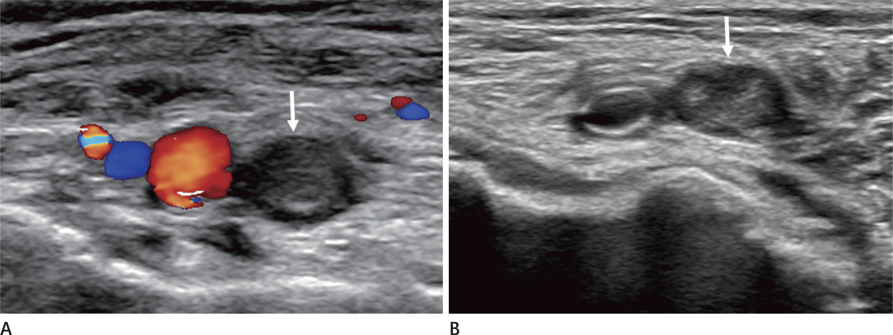
Ultrasonography of an 89-year-old female with acute deep vein thrombosis in her right leg. A. In color Doppler ultrasonography, there is no flow through the popliteal vein (arrow). B. This vein does not collapse upon compression (arrow).

Fig. 2.
Computed tomography venography of an 84-year-old male patient. There is an abnormal dilated left common femoral vein with hypodense filling defects in the lumen and subtle vascular wall enhancement (arrow). These findings are compatible with an acute stage of deep vein thrombosis. Combined subcutaneous edema is noted.

Fig. 3.
Computed tomography venography of a 71-year-old female patient with a previous history of May-Thurner syndrome 9 years previously. The stent of the left common iliac vein, placed 9 years ago, is occluded (A, arrow). The left external iliac vein is in a near complete obliteration state (B, arrow) and multiple collateral vessels are noted in the subcutaneous layer of anterior lower abdominal wall and pelvic cavity (C, arrows).

Fig. 4.
Acute and chronic DVT in conventional leg ascending venography. A. A 66-year-old female with an acute DVT in her left leg. Multiple filling defects are noted in the femoral and popliteal veins outlining the dilated veins. Femoral vein duplication (arrows) is noted. B. A 63-year-old male with chronic DVT in her right leg. The obliteration of femoral vein with multiple collateral vessels is noted. DVT = deep vein thrombosis

Fig. 5.
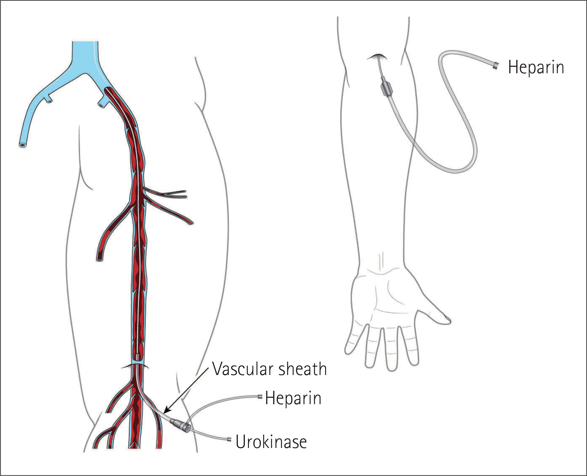
Illustration of the catheter directed thrombolysis. The vascular sheath is inserted via the popliteal vein. Through this vascular sheath, a 5-Fr multi-sidehole infusion catheter is inserted with the tip embedded in the thrombus. Through this infusion catheter, a thrombolytic agent is infused. An anticoagulant is infused via the both of side arm of the vascular sheath and other peripheral intravenous line.

Table 1.
Risk Factors of the Development of Deep Vein Thrombosis


 PDF
PDF ePub
ePub Citation
 Print
Print


 XML Download