

 PDF
PDF ePub
ePub Citation
 Print
Print


INTRODUCTION
Zinner's syndrome is a rare congenital abnormality of mesonephric duct. Unilateral renal agenesis, ipsilateral seminal vesicle cyst, and ipsilateral ejaculatory duct obstruction are the triad of maldevelopment of mesonephric duct consisting Zinner's syndrome. In embryogenesis, urinary tracts and genital organs develop from mesonephric duct, called urogenital ridge. If there is any signal interference or mistake, congenital anomaly can develop in the urinary tracts and also genital organs including internal genitalia such as testis and prostate (1).
CASE REPORT
A 23-year-old male presented with history of urinary retention and dysuria. There was microscopic hematuria without proteinuria, pyuria or malignant cells.
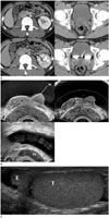
Computed tomography (CT) showed renal agenesis of right kidney with compensatory hypertrophy of the normally situated left kidney and a cystic mass in the right seminal vesicle. Left kidney had homogeneous enhancement without perfusion defects in nephrographic phase and good excretory function (Fig. 1A). Patient's serum blood urea nitrogen and creatine level were within normal range, suggesting well-functioning left kidney.
In addition, there was a bi-lobed filling defect in the bladder trigone on excretory phase CT scan (Fig. 1A), showing the same echogenicity with prostate on transrectal ultrasonography (TRUS). The overlying bladder mucosal layer was intact. The cystic lesion in the right seminal vesicle, previously noted on CT scan was also seen on TRUS as dilated tubular structure (Fig. 1B). At the same time, scrotal ultrasonography (US) using a linear probe revealed an additional testis at the bottom of normal right testis in the scrotum (Fig. 1C).
Semen analysis showed ejaculate volume of 2.9 mL (normal ejaculatory volume, > 1.5 mL), mild oligospermia with sperm count of 4.0 × 10⁶/mL (normal sperm count, > 15 × 106/mL) and pH 9 (normal semen pH 7.2–7.8). If there is an obstruction in the ejaculatory duct, oligospermia or azoospermia can be found in semen analysis. Furthermore, increased semen pH means decreased fructose level in semen, meaning obstruction at the ejaculatory duct (2). Consequently, ejaculatory duct obstruction is highly suspicious in our patient.
To summarize, CT and sonographic findings showed the agenesis of right kidney with an ipsilateral seminal vesicle cyst, probably caused by the ejaculatory duct obstruction. Even though the patient had not undergone histologic confirmation, ectopic prostate and triorchidism are strongly suspicious based on their typical locations and similar echogenicities with normal testis and prostate on US as well. In short, our patient was Zinner's syndrome, which is a developmental anomaly of the mesonephric duct with other mesonephric duct associated genital anomalies, ectopic prostate and triorchidism.
DISCUSSION
Zinner's syndrome is a rare congenital malformation originates from the mesonephric duct. Most patients are asymptomatic until the third or fourth decade of life and often manifest symptoms during the period of high sexual or reproductive activity (3).
The close relationship between genital and urinary tract in embryology explains the triad of Zinner's syndrome. The urogenital system develops from the urogenital ridge and the cloaca. The urogenital ridge develops into three sets of tubular nephric structures, i.e., pronephros, mesonephros, and metanephros. It is also associated with gonadal development. The cloaca, which is the terminal portion of the hindgut develops urogenital sinus and anal canal. The mesonephric duct, known as Wolffian duct, develops into hemitrigone, bladder neck and internal genitalia such as urethra, seminal vesicle, vas deferens, ejaculatory ducts, epididymis, and appendix epididymis. The ureteric buds develop from the caudal portion of the mesonephric duct and secrete growth factors and proliferates, fusing with the metanephric blastema. The ureteric buds and metanephric blastemas consist the metanephric duct and become the primitive kidneys and urinary tracts (4).
Disturbance in any of these developments causes congenital anomalies of the urinary tracts or male internal genitalia. Failure of fusion of the ureteric bud with the metanephric blastema causes renal agenesis or renal hypoplasia. Simultaneous failure of the ureteric bud to separate from the lower part of mesonephric duct leads to atresia of ejaculatory ducts and obstruction of the seminal vesicles, resulting in cystic dilatation (3). When the cystic dilatation becomes significant size, lower urinary tract symptoms including dysuria can occur due to its mass effect (4). The symptoms our patient complained-urinary retention and dysuria-could be caused by this mechanism.
In our case, though it was not confirmed by pathology, the patient is assumed to have the ectopic prostate and triorchidism. The ectopic prostate is another result from mesonephric duct anomaly which makes internal genitalia and bladder trigone. It usually involves bladder trigone or ureteral orifices (5) or rarely outside the urinary tract (6). Other entities like submucosal neoplasms of the bladder can be considered. Mesenchymal tumor arises in the bladder wall and most common type is leiomyoma, which usually presents as a homogeneous hypoechoic mass with few blood vessels on color Doppler US (7). Triorchidism can also be explained as a result of mesonephric duct anomaly. The urogenital ridge differentiates into medial genital ridge and a lateral nephrogenic ridge. The medial genital ridge is responsible for development of internal genitalia. If there is any interruption or degeneration in the mesonephric duct, there can be anomalies in internal genitalia such as polyorchidism. Triorchidism is most common form of polyorchidism (8) and testicular duplication may develop due to the duplication of the genital ridge or longitudinal or transverse division of it (9). Ectopic prostate and triorchidism are rare congenital anomalies and this is the first time to report the patient with Zinner's syndrome has these rare congenital anomalies.
When patients present with congenital anomalies of urinary tract, radiologist should suggest evaluating not only urinary tracts but also genitalia because imaging studies play an important role to discover the triad of Zinner's syndrome and associated anomalies. Image studies can reveal other obscured congenital anomalies and furthermore, can suggest the reason of clinical symptoms such as subfertility or urinary symptoms. If patient underwent abdominal surgery, these congenital anomalies can cause confusion to surgeons. Understanding embryologic association of the urinary tract and genitalia is the key point of diagnosing congenital anomalies of the mesonephric duct and avoiding unnecessary invasive procedures or surgery. In conclusion, we have described an extremely rare case of Zinner's syndrome with other mesonephric duct associated congenital anomalies.
 XML Download