

 PDF
PDF ePub
ePub Citation
 Print
Print


INTRODUCTION
When performing interventional procedure such as transarterial chemoembolization (TACE), we encounter lots of anatomic variations of celiac axis and hepatic arteries. To have better outcomes and prevent iatrogenic complications, it is essential to be aware of these variations.
Previously published reports have demonstrated that the incidence of classical trifurcation pattern of celiac axis was 70.8% (1); the accessory left gastric artery (LGA) arising from the left hepatic artery (LHA) was 11.1–14.2% (23); the accessory LHA arising from the LGA was 6.25–17.9% (13); and the replaced LHA from LGA in 6.25% (1). The first report was based on visual inspections during surgical operations, angiograms and magnetic resonance imaging studies. Other two reports were based on visual inspections during autopsies and on angiograms, respectively. However, none of these reports had the analysis using computed tomography (CT), which is considered more effective to precisely display the anatomical detail of blood vessels.
We recently encountered a rare case of the entire LGA arising from the LHA in a 62-year-old man during TACE for recurred hepatocellular carcinoma (HCC), and verified this anatomical variant by 3-phase liver CT with 3-dimensional volume rendering reconstruction as well as angiography.
CASE REPORT
A 62-year-old male, diagnosed with HCC seven years ago and a hepatitis B carrier, underwent 3-phase liver CT during his regular surveillance. Previously, he had received TACE two times and radiofrequency ablation five times in a row, and had not experienced surgical operations for HCC. For the following six months, he remained free of recurrence. However, the CT image showed a newly developed mass with typical features of HCC. The mass was measured at 1.4 cm and located in the segment II of the left hepatic lobe. The patient was referred to the interventional radiologist for TACE.
The right common femoral artery was accessed percutaneously, and celiac angiography using 5-Fr catheter was carried out. The splenic artery and the common hepatic artery (CHA) were arising from the celiac axis without abnormality. However, the LGA did not appear to be in the celiac axis, its most common origin. Subsequent common hepatic arteriogram revealed a vessel with ascending course originating from proximal to the umbilical point of the LHA, and running along the gastrohepatic ligament. At first, we considered it as an accessory LGA from the LHA since the course was about the same as that of an accessory LGA. In addition, there was still the possible presence of the LGA from other mother vessels such as aorta.
After cone-beam CT was taken, superselective arteriograms of the tumor-supplying vessels were obtained using 2-Fr microcatheter. Chemoembolization using doxorubicin was also performed via A2 sub-branch from the LHA passing by the originating point of the LGA.
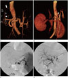
After the procedure, we reviewed all of the past images obtained by 3-phase liver CT with volume rendering reconstruction and angiography. We checked again the fact that the patient had not experienced any kinds of surgical operations at all. The celiac trunk showed an aberrant branching pattern (Fig. 1A); it bifurcated into the CHA and the splenic artery. The course of the CHA was normal. However, the LGA arose from the LHA and was running through the fissure for ligamentum venosum, then along the gastrohepatic ligament. The course was almost the same as that of a usual accessory LGA, which is known to originate commonly from proximal to the umbilical point of LHA (3). The diameter of the LGA was as large as the main LHA, and larger than a usual accessory LGA. Relatively narrower spectrum of the lesser curvature from the left side of the stomach was supplied by the LGA. The right gastric artery (RGA) was thicker and longer than a usual RGA and was supplying a broad spectrum of the lesser curvature from the right side of the stomach.
Because the patient underwent three times of TACE, we could not exclude the possible degeneration of additional features of the LGA which existed in the past. Therefore, we thoroughly analyzed initial CT and angiographic images one by one, and found no any other feature of the LGA from the celiac axis, aorta and other possible mother arteries (Fig. 1B-D). Against this back-drop, we concluded that the LGA was an entirely replaced LGA from the LHA.
DISCUSSION
A case of a completely replaced LGA arising from the LHA has not been documented in the literature regarding anatomic variations of celiac axis and its branching vessels (245). In 1978, Naidich et al. found the case of the replaced LGA arising from the LHA in two of 500 reviewed celiac angiograms (0.4%) in his classic article about the origin of LGA without CT images (6). In comparison with the report of Naidich which covered limited selective angiographic studies of the celiac axis, we confirmed the LGA from the LHA as the solitary main vessel without any accessory features by using CT studies including the volume rendering techniques. CT angiography is considered better to precisely display the anatomical detail of blood vessels, and it is reported that 70% of accessory LGAs can be diagnosed at the early phase of multidetector CT even with a slice thickness of 5 mm (7).
We are open to the possibility that our imaging findings can be interpreted in two other ways. First, the replaced LGA arises from the proper hepatic artery, then the LHA arises from the LGA. Second, the common trunk of LHA and LGA arises from the proper hepatic artery, then it bifurcates into the LHA and the replaced LGA.
In addition, it is noteworthy that during the review of the images (Fig. 1A) we found an unusual communication between the RGA and the gastroduodenal artery.
Patients who underwent TACE can suffer upper gastrointestinal complications such as bleeding, gastritis, and ulceration caused by infused embolic agents into the gastric arteries, especially when anatomic variants exist. It has documented that upper gastrointestinal bleeding occurred in 3% (range 0–22%) of 2593 patients in 23 trials (8). To reduce the risk of these complications, it is significant to be aware of anatomical variations and possible communications of hepatic and gastric arteries, prior to performing any procedures.
Furthermore, the catheterization of LGA has a clinical importance in controlling of acute gastrointestinal hemorrhage. It is known that the LGA supplies 85% of all angiographically-documented upper gastrointestinal bleeding sites (9). The empiric embolization of the LGA appears to be helpful when CT or angiography fails to reveal active bleeding foci (10).
The accessory LGA arising from the LHA is one of relatively common anatomical variations, but the case of the replaced LGA arising from hepatic arteries has rarely been documented. This is the first report that confirmed the completely replaced LGA from LHA by both CT and angiographic images. We believe the awareness of the possible presence of the entirely replaced LGA without any other additional accessory features can help interventional radiologists and surgeons in planning various procedures and surgical operations such as TACE, gastric artery embolization, gastrectomy and hepatic transplantation.
 XML Download