

 PDF
PDF ePub
ePub Citation
 Print
Print


INTRODUCTION
Dermatofibrosarcoma protuberans (DFSP) is a rare, malignant cutaneous soft tissue tumor. DFSP is locally aggressive and can recur after surgical resection. It commonly occurs on the trunk and extremities, but rarely involves the breast (12). We report here the ultrasonographic and histopathologic findings of a rare case of DFSP in the breast.
CASE REPORT
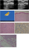
A 43-year-old woman with no previous medical history, presented with a growing palpable mass in the left breast that had been present for 2 years, with accompanying itching and pain. Physical examination showed a firm, non-tender mass with overlying erythematous skin changes on the left upper inner breast. Mammography revealed no abnormal lesions. Breast ultrasonography shows a 1.4 × 1.2 × 0.3 cm oval shaped low echoic mass in the dermal layer, with surrounding increased subcutaneous fat echogenicity at the 10 o'clock direction of the left breast (Fig. 1A). Primary diagnosis was benign dermal lesion such as infected epidermal cyst. However, an excision biopsy was performed and pathology revealed DFSP. So, a subsequent wide excision was carried out. On gross specimen, about 1.5 × 0.4 cm nodular mass locates in the dermal layer with infiltration into subcutaneous fat (arrow) (Fig. 1B). On histopathologic examination, the mammary skin shows a highly cellular spindle cell tumor proliferating in the dermal layer, just beneath the thinned epidermis (Fig. 1C). Fibroblastic tumor cells are growing closely packed and radially in a so-called storiform pattern (Fig. 1D). The tumor cells are diffusely immunoreactive for anti-CD34 antibody, a diagnostic marker for dermatofibrosarcoma protuberans (Fig. 1E). These tumor cells infiltrate into subcutaneous fat, producing a characteristic “honeycomb pattern” (arrow) (Fig. 1F). The patient was followed up for 5 years using ultrasonography, and did not exhibit any signs of recurrence.
DISCUSSION
DFSP is a rare, locally aggressive cutaneous soft tissue tumor having intermediate malignancy, with the most common being mesenchymal superficial malignancy. DFSP arises from the dermis, but some lesions are known to infiltrate into deeper layers. It can recur after surgical resection, but distant metastasis rarely occurs (12). The incidence of DFSP was reported to be about 5 cases per 1 million persons per year (3). DFSP accounts for approximately 1.8% of all soft tissue sarcomas (2), and is usually manifested as a slowly growing superficial red or bluish-red plaque (2). It commonly occurs on the trunk, extremities, and in the head and neck areas (4); involvement of DFSP in the skin of breast is rare. Several previous literatures have reported its imaging findings (5678).
Fukushima et al. (5) reported a case of DFSP in the breast, which was hypoechoic with irregular internal echogenicity on ultrasound and initially misdiagnosed as a primary breast lesion such as fibroadenoma or phyllodes tumor. Parajuly and Peng (6) reported four cases of DFSP in the breast. In three of these cases, ultrasonography showed oval-shaped, hypoechoic mass originating from the dermis of the breast, with an internal irregular echogenicity. However, one case was misdiagnosed as primary breast lesion due to location of the mass in the mammary tissue. In our case, the mass was located in the dermal layer above the subcutaneous fat layer, and this feature differentiated the mass from a primary breast lesion. Thus, similar to our case, by using ultrasonography, DFSP with superficial location could be differentiated from primary breast lesion.
In the present case, as the mass was located in the dermal layer, benign dermal lesion (such as infected epidermal cyst) was suspected, considering the size, margin and parallel orientation. However, histopathologic examination confirmed a diagnosis of DFSP. Retrospectively, different ultrasonographic features were noted in the current case of DFSP in the breast. Increased subcutaneous fat echogenicity around the mass was observed, and it was more prominent than the mass size. Moreover, histopathologic examination showed the tumor cells of DFSP infiltrate into deep dermis and subcutaneous fat, producing a characteristic “honeycomb pattern.” The honeycomb pattern is an essential histologic characteristic of DFSP, which is due to infiltration of tumor cells into the subcutaneous tissue (9). Therefore, ultrasonographic features of increased subcutaneous fat echogenicity around the mass correlated well with histopathologic features of infiltration of tumor cells into the subcutaneous fat (the honeycomb pattern).
Lee et al. (7) reported five cases of DFSP in the breast and reviewed five previously reported cases. In this study, all tumors were oval shaped, parallel in orientation, and predominantly circumscribed with some microlobulation. An incomplete thin hypoechoic rim along the deep margin of the tumor was noted in three cases. One case of DFSP in the breast has also been reported by Kang et al. (8). In the case, ultrasonography showed lobulated and partially irregular margin of the tumor, and the histopathologic examination showed characteristic diffuse infiltration of fibroblast between fat cells in the subcutaneous tissue. Shin et al. (10) reported ultrasonographic findings of four DFSP located in the back area. These radiologic features included pseudopodia like protrusion of the tumor, and a focally disrupted thin hypoechoic rim by the tumor's protrusion; these features correlated with the histopathologic findings of infiltration of tumor cells into subcutaneous fat. However, no previous literature has reported increased subcutaneous fat echogenicity around the mass (which is more prominent than the mass size) as an ultrasonographic feature of DFSP in the breast. This ultrasonographic feature reflects an infiltration of tumor cells into subcutaneous fat.
In conclusion, the involvement of breast in DFSP is rare, making it difficult to diagnose. Occasionally, due to its infiltrative nature, some cases involve mammary tissue and simulate primary breast lesions. However, DFSP of breast is usually located at the superficial layer (dermal layer or subcutaneous fat layer). Therefore, ultrasonography helps in differentiating it from primary breast lesion. The present case illustrates DFSP in the breast, wherein ultrasonography enabled the differentiation of DFSP from primary breast lesion. Also, in this case, a unique ultrasonographic feature of increased subcutaneous fat echogenicity around the mass was noted. This finding correlated with the pathologic finding of infiltration of tumor cells into subcutaneous fat. On breast ultrasonography, if low echoic mass at the superficial layer (dermal layer or subcutaneous fat layer) with prominent increased subcutaneous fat echogenicity around the mass is noted, we can include DFSP as a differential diagnosis, along with other benign lesions. This can help in proper management decision, such as biopsy or surgical excision, to be made.
 XML Download