

 PDF
PDF ePub
ePub Citation
 Print
Print


INTRODUCTION
Bezoars are the accumulation of ingested materials. Different types of bezoars can occur in human and their name reflects the ingested material, such as vegetable or fruit fiber (phytobezoar) and milk curd (lactobezoar). The most common type of bezoar is trichobezoar, which is made of hairs (12).
Trichobezoars are formed in the stomach but sometimes may
pass through the pylorus into small intestines (Rapunzel Syndrome). The name came after Grimm Brothers' fairy tale in 1812 about a 12-year-old maiden who lowered her long hair from a tower to let her prince climbing up the tower and rescue her. The syndrome was discovered for the first time by Vaughan in 1968 and since then approximately 30 cases have been reported in the literature (1234).
CASE REPORT
A 9-year-old girl complained about intermittent abdominal pain that subsided after vomiting since 1 year ago. She had a poor apetite and early satiety. There was also a lump in her abdominal. Patient had a history of Trichotillomania and Trichophagia since the age of 2 years and never received psychiatric treatments before.
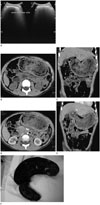
Ultrasonography (US) was performed using 3.5 MHz transducer ultrasound (MyLab™ClassC; Esaote, Genoa, Italy). Abdominal US on the mass revealed an arc-like hyperechoic curvilinear with posterior acoustic shadow (Fig. 1A). Computed tomography (CT) examination was performed using a 64-channel multi detector CT (Somatom Definition AS; Siemens, Erlangen, Germany). On CT, there was a heterogenous intragastric mass that extended to superior part of duodenum with air bubbles within and there was no enhancement in the mass with intravenous contrast administration (Fig. 1B-E).
The patient underwent a laparotomy and a hair ball, which weighed about 1 kg, was removed. It had a perfect cast of the stomach, pylorus, and superior part of duodenum (Fig. 1F). After the surgery, psychiatric consultation was scheduled and the patient was discharged home 6 days after the surgery.
DISCUSSION
Trichobezoars are associated with psychiatric disorders including Trichotillomania and Trichophagia. Most of cases have been reported in young females and in countries where women traditionally have long hair (145).
The ingested hairs escape from peristaltic because of their slippery surface and detained in the gastric mucosa folds. Gastric peristalsis causes hairs to be enmeshed into a ball shape. The hairball becomes even more matted together and casts the shape of the stomach, as a solid mass. Because of the hold up by the pylorus and the churning action of the stomach, the hairball usually located in the stomach (135).
The gold standard for diagnosis trichobezoar is upper gastrointestinal endoscopy (24). Even though upper gastrointestinal is the gold standard diagnosis for trichobezoar, it may not prove the presence of a co-existing Rapunzel Syndrome.
The diagnosis of Rapunzel Syndrome depends on the use of CT and intraoperative finding both. However, there are various diagnostic criteria to describe Rapunzel Syndrome. First is a trichobezoar with a tail extend up to ileocecal junction; other criteria is a trichobezoar with a long tail extend up to jejunum or beyond; or as a trichobezoar of any size which can cause intestinal obstruction (13).
The sonographic appearance of trichobezoar was a hyperechoic curvilinear (arc-like) with acoustic shadow. However, there were difficulties in revealing multiple bezoars and unable to locate bezoars that far from the abdominal surface (67).
CT is a superior to other radiologic tools for trichobezoars diagnosis and excludes differential diagnosis in patients with intestinal obstruction. Hairballs appear as non-enhanced, well-described, heterogenous, intraluminal mass with mottled appearance. The heterogenous and mottled appearances come from different densities of its material, including food debris and air-bubble (27).
Trichobezoar and Rapunzel Syndrome are bizarre, uncommon diseases. Radiologists should be able to recognize and diagnose them precisely. Imaging plays an important role to make a diagnosis and exclude the differential diagnosis in young women with symptoms of intestinal obstruction. In order to avoid complications, an early and accurate diagnosis is important, which is able to be made by imaging.
 XML Download