

 PDF
PDF ePub
ePub Citation
 Print
Print


INTRODUCTION
Retroperitoneal extraskeletal osteosarcoma is rare malignant mesenchymal tumor with an osteoid component without contact to bony structure(s) (1). On computed tomography (CT), soft tissue tumor(s) show various levels of ossified matrix; therefore, it is crucial to detect calcification to narrow the differential diagnosis. However, approximately 50% of extraskeletal osteosarcomas do not exhibit gross calcification on imaging studies. Therefore, it is difficult for radiologists to diagnose extraskeletal osteosarcoma arising in the retroperitoneal space without calcification on CT scan. We report a case of retroperitoneal extraskeletal osteosarcoma, without calcification on CT scan, mimicking a pancreatic tumor, and describe its imaging features with ancillary clinical information to facilitate diagnosis.
CASE REPORT
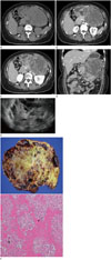
A 44-year-old woman presented with a 10-month history of a palpable mass in the abdomen. Laboratory examinations were unremarkable except for elevation of alkaline phosphatase level [159 IU (normal range 40–130 IU)]. To evaluate her symptoms, abdominal CT was performed with contrast enhancement, which revealed a large (approximately 16.5 cm) soft tissue mass with lobulated contours in left side of the retroperitoneum (Fig. 1). There was no definite radiopaque lesion suggesting calcification or fat attenuation within the mass on the precontrast scan (Fig. 1A). On the arterial phase scan, the mass abutted the body of the pancreas body and obscured the pancreas body and tail. Obliteration of the splenic vein was also noted on the portal venous phase (Fig. 1C, D). The enhancement pattern of the mass was heterogeneous on the arterial and portal phases (Fig. 1B, C). The peripheral portion of the mass exhibited persistent enhancement, while the central portion exhibited a low-density lesion without contrast enhancement, suggesting cystic or necrotic change(s) (Fig. 1C, D). It was assumed that the origin of the mass was in the pancreas and made impression with solid and cystic masses of the pancreas, such as acinar cell carcinoma and solid pseudopapillary neoplasm. On endoscopic ultrasonography, the echotexture of the mass was heterogeneous and mixed with solid and cystic components (Fig. 1E). Endoscopic ultrasonography also suggested that the mass did not originate from the stomach, similar to a gastrointestinal stromal tumor, because layers of the gastric wall were maintained.
Surgical resection was the chosen treatment option. The mass adhered to the stomach and appeared to originate from the pancreas. Grossly, the tumor was a cystic and solid mass with lobulated contours (Fig. 1F). Engorged vessels in the tumor were noted. After dissection of the adhesion between the mass, stomach, and celiac trunk, the mass was resected with the distal pancreas. Microscopic examination revealed deposition of a network of irregular, eosinophilic, glassy osteoids with interspersed cellular stroma composed of spindle or plump cells (Fig. 1F). The pathological diagnosis was extraskeletal osteosarcoma. Positron emission tomography CT performed before surgery revealed no other hot uptake lesions except for the retroperitoneal mass. It was therefore concluded that the retroperitoneal extraskeleta osteosarcoma was the primary lesion.
DISCUSSION
Extraskeletal osteosarcoma is rare malignant mesenchymal tumor consisting of osteoid tumor cells without bony involvement (12). It accounts for approximately 4% of all osteosarcomas and 1% of all soft tissue sarcomas (12). Primary osteosarcoma of the bone occurs mostly in the first decade of life; however, extraskeletal osteosarcomas occur in adults 50 to 70 years of age (13). The most common sites of tumor occurrence are the lower extremities (47%), upper extremities (20%), retroperitoneum (17%), and trunk/other (10%) (1234); men are more frequently affected than women (1). Although elevation of serum alkaline phosphatase levels is a common finding in osteosarcomas, including retroperitoneal extraskeletal osteosarcomas, in other conditions also exhibiting retroperitoneal mass(es), it is not prone to elevation (3). Pathogenesis is unclear; however, a few risk factors have been reported including previous radiation exposure or trauma. Other studies have proposed that myositis ossificans may be a precursor of extraskeletal osteosarcoma (25). Retroperitoneal extraskeletal osteosarcoma appears as various amorphous shapes or intense calcifications (4). On CT scan, matrix ossification or calcification could be present in approximately one-half of primary tumors; moreover, calcification may represent tumor progression (1). Extraskeletal osteosarcoma has been reported to exhibit a pseudocapsule and no contact with adjacent bony structures on CT (1). CT reveals a soft tissue mass often displacing fat planes, with variable degrees of matrix mineralization. Nonmineralized soft tissue exhibits attenuation consistent with that of muscle on CT. On post-contrast CT, extraskeletal osteosarcomas are usually heterogeneously enhanced depending on the degree of necrosis and hemorrhage. The tumors can directly invade adjacent structures (12345).
The radiological differential diagnosis of retroperitoneal extraskeletal osteosarcoma mimicking pancreatic tumor includes pancreatic solid and cystic masses, such as solid pseudopapillary tumor, invasive intraductal papillary mucinous neoplasm, and acinar cell carcinoma (67). Solid pseudopapillary tumors have smooth margins, with various levels of solid and cystic components, and occurs mostly in young women (67). Invasive intraductal papillary mucinous neoplasm, especially the branched duct type, also appears as a solid and cystic mass but does not exhibit calcification and, on magnetic resonance cholangiopancreatography reveals communication with the pancreatic duct (6). Acinar cell carcinoma is a rare epithelial pancreatic tumor that is typically well-defined and homogeneously hypovascular; however, when it is large, it may exhibit various levels of cystic portions due to necrosis (7). Nevertheless, it is difficult to predict the specific pathological type of the retroperitoneal masses on imaging studies (8). On the other hand, as reported, elevation of serum alkaline phosphatase level could be helpful in the differential diagnosis of extraskeletal osteosarcoma (3).
The treatment of extraskeletal osteosarcoma is variable, including radical excision only, adjuvant or neoadjuvant chemotherapy with excision, and radiotherapy (9). The prognosis for extraskeletal osteosarcoma is not favorable, with a reported 5-year survival rate of approximately 60% due to metastasis and recurrence (approximately 50%) (9). The most important factors in the prognosis of extraskeletal osteosarcoma are the size and location of the tumor. Smaller tumor sizes (< 5 cm) and more superficial locations, such as the extremities or trunk, rather than the retroperitoneal or intra-abdominal space, make complete resection possible (9).
We reported a case of extraskeletal osteosarcoma in the retroperitoneal space without calcification mimicking a pancreatic tumor on CT. Because of its varying morphology on imaging studies, it is difficult to predict a diagnosis from imaging before pathological confirmation. Additionally, tumor invasion of the pancreas was a confusing factor in determining the origin of the mass. We suggest that if a large retroperitoneal heterogeneous mass is encountered, even without calcification on CT, in addition to careful review of clinical characteristic, such as elevation of serum alkaline phosphatase levels, retroperitoneal extraskeletal osteosarcoma should be considered in the differential diagnosis.
 XML Download