Abstract
Purpose
To compare the diagnostic performance of magnetic resonance (MR) sequences for the evaluation of cerebral venous sinus thrombosis (CVST) during followup examinations.
Materials and Methods
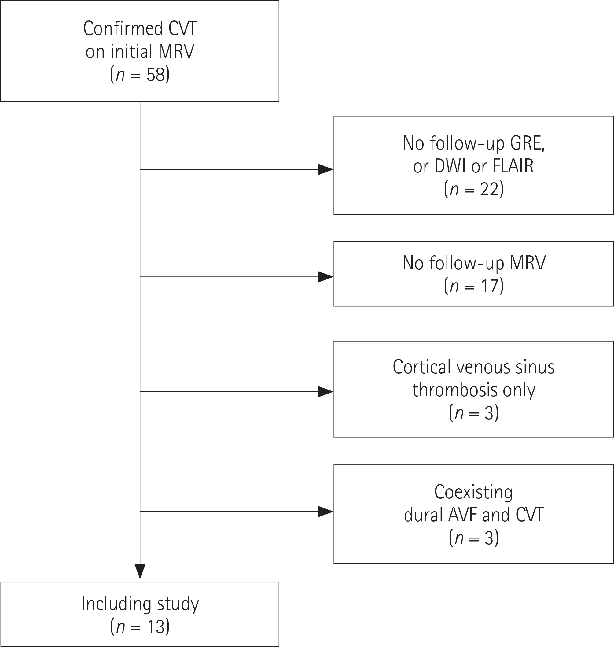
Thirteen cases that were confirmed to be CVST between January 2006 and March 2016 were included in this study. Two neuroradiologists independently examined each initial and follow-up MR sequence image in random order.
Results
Gadolinium-enhanced T1-weighted imaging (Gd-enhanced T1WI) was the most sensitive sequence for the detection of CVST in the initial and follow-up MR examinations (82% and 55.3%, respectively). Among the non-enhanced MR sequences of the initial examination, gradient-recalled echo was the most sensitive (77.4%), fluid-attenuated inversion recovery (FLAIR) had low sensitivity (34.4%). The overall diagnostic performances of all MR sequences except for FLAIR decreased during the follow-up. FLAIR was the most sensitive during follow-up, and was also the only sequence with increased sensitivity during follow-up (from 34.4% to 55.6%).
Conclusion
Gd-enhanced T1WI had the best diagnostic performance for CVST in both initial and follow-up MR examinations. Therefore, it is reasonable to use Gd-enhanced T1WI to evaluate CVST during follow-up examinations. However, for patients who cannot tolerate MR contrast agents, the use of FLAIR to assess the remaining CVST during the follow-up may be helpful.
Go to : 
References
3. Coutinho JM, Zuurbier SM, Aramideh M, Stam J. The incidence of cerebral venous thrombosis: a cross-sectional study. Stroke. 2012; 43:3375–3377.
4. Devasagayam S, Wyatt B, Leyden J, Kleinig T. Cerebral venous sinus thrombosis incidence is higher than previously thought: a retrospective population-based study. Stroke. 2016; 47:2180–2182.
5. Tatlisumak T, Jood K, Putaala J. Cerebral venous thrombosis: epidemiology in change. Stroke. 2016; 47:2169–2170.
6. Gulati D, Strbian D, Sundararajan S. Cerebral venous thrombosis: diagnosis and management. Stroke. 2014; 45:e16–e18.
7. Ferro JM, Canhão P. Cerebral venous sinus thrombosis: update on diagnosis and management. Curr Cardiol Rep. 2014; 16:523.

8. Filippidis A, Kapsalaki E, Patramani G, Fountas KN. Cerebral venous sinus thrombosis: review of the demographics, pathophysiology, current diagnosis, and treatment. Neurosurg Focus. 2009; 27:E3.

9. Puig J, Pedraza S, Blasco G, Serena J. [Review of the neuroradiological diagnosis of cerebral venous thrombosis]. Radiologia. 2009; 51:351–361.
10. Idbaih A, Boukobza M, Crassard I, Porcher R, Bousser MG, Chabriat H. MRI of clot in cerebral venous thrombosis: high diagnostic value of susceptibility-weighted images. Stroke. 2006; 37:991–995.
11. Sajjad Z. MRI and MRV in cerebral venous thrombosis. J Pak Med Assoc. 2006; 56:523–526.
12. Moreno-Ramos MD, Rodríguez-Romero R, Piñero-González de la Peña P, Rodríguez-Uranga J, Monreal-Monsalve C. [Noninvasive diagnosis of cerebral venous thrombosis]. Radiologia. 2006; 48:79–86.
13. Leach JL, Fortuna RB, Jones BV, Gaskill-Shipley MF. Imaging of cerebral venous thrombosis: current techniques, spectrum of findings, and diagnostic pitfalls. Radiographics. 2006; 26(Suppl 1):S19–S41. ; discussion S42-S43.

14. Leach JL, Strub WM, Gaskill-Shipley MF. Cerebral venous thrombus signal intensity and susceptibility effects on gradient recalled-echo MR imaging. AJNR Am J Neuroradiol. 2007; 28:940–945.
15. Dormont D, Anxionnat R, Evrard S, Louaille C, Chiras J, Marsault C. [MRI in cerebral venous thrombosis]. J Neuroradial. 1994; 21:81–99.
16. Hinman JM, Provenzale JM. Hypointense thrombus on T2-weighted MR imaging: a potential pitfall in the diagnosis of dural sinus thrombosis. Eur J Radiol. 2002; 41:147–152.

17. Bidar F, Faeghi F, Ghorbani A. Assessment of cerebral venous sinus thrombosis using T2 (∗)-weighted gradient echo magnetic resonance imaging sequences. Iran J Neurol. 2016; 15:96–99.
18. Ihn YK, Jung WS, Hwang SS. The value of T2∗-weighted gradient-echo MRI for the diagnosis of cerebral venous sinus thrombosis. Clin Imaging. 2013; 37:446–450.

19. Selim M, Fink J, Linfante I, Kumar S, Schlaug G, Caplan LR. Diagnosis of cerebral venous thrombosis with echo-planar T2∗-weighted magnetic resonance imaging. Arch Neurol. 2002; 59:1021–1026.

20. Meckel S, Reisinger C, Bremerich J, Damm D, Wolbers M, Engelter S, et al. Cerebral venous thrombosis: diagnostic accuracy of combined, dynamic and static, contrast-enhanced 4D MR venography. AJNR Am J Neuroradiol. 2010; 31:527–535.

21. Sadigh G, Mullins ME, Saindane AM. Diagnostic performance of MRI sequences for evaluation of dural venous sinus thrombosis. AJR Am J Roentgenol. 2016; 206:1298–1306.

22. Favrole P, Guichard JP, Crassard I, Bousser MG, Chabriat H. Diffusion-weighted imaging of intravascular clots in cerebral venous thrombosis. Stroke. 2004; 35:99–103.

23. Chu K, Kang DW, Yoon BW, Roh JK. Diffusion-weighted magnetic resonance in cerebral venous thrombosis. Arch Neurol. 2001; 58:1569–1576.

24. Walecki J, Mruk B, Nawrocka-Laskus E, Piliszek A, Przelas-kowski A, Sklinda K. Neuroimaging of cerebral venous thrombosis (CVT) – old dilemma and the new diagnostic methods. Pol J Radiol. 2015; 80:368–373.

Go to : 
 | Fig. 1.Study flow chart. AVF = arteriovenous fistula, CVT = cerebral venous thrombosis, DWI = diffusion-weighted imaging, FLAIR = fluid-attenuated inversion recovery, GRE = gradient-recalled echo, MRV = magnetic resonance venography |
 | Fig. 2.Images of a 51-year-old man with acute left transverse sinus thrombosis (3 days). Note the susceptibility artifact, which is low signal and blooming (white arrows) on gradient-recalled echo imaging (A) and the empty delta sign (black arrows) in the left transverse sinus on Gd-enhanced T1-weighted imaging (B). No signal change is observed in the left transverse sinus (black arrows) compared to the normal right transverse sinus on fluid-attenuated inversion recovery (C). No diffusion restriction is observed in the left transverse sinus (white arrows) on diffusion-weighted imaging (D). Dural venous sinus thrombosis is confirmed as the filling defect in left transverse and sigmoid sinuses on gadolinium-enhanced 4 dimensional magnetic resonance venography (E). |
 | Fig. 2.(continued) Images of a 51-year-old man with left transverse sinus thrombosis during follow-up (12 days). The patient underwent followup brain magnetic resonance imaging including MRV 12 days after the first examination due to aggravation of the headache. Note the susceptibility artifact on gradient-recalled echo (F) and typical empty delta sign in the left transverse sinus on Gd-enhanced gadolinium-enhanced T1-weighted imaging (G) are not observed (black arrows). The high signal intensity (black arrows) on fluid-attenuated inversion recovery (H), and high signal intensity indicating diffusion restriction (black arrows) in the left transverse sinus on diffusion-weighted imaging (I). Dural venous sinus thrombosis is confirmed as the filling defect in the left transverse and sigmoid sinuses on follow-up gadolinium-enhanced 4 dimensional MRV (J). MRV = magnetic resonance venography |
Table 1.
Clinical Characteristics of 13 Patients with Dural Venous Sinus Thrombosis
Complete = complete recanalization, Duration = days from symptom onset to first MRI, F = female, F/U = follow-up, IS = inferior sagittal sinus, LS = lef sigmoid sinus, LT = left transverse sinus, M = male, MRI = magnetic resonance imaging, No = no recanalization, Partial = partial recanalization, RS = righ sigmoid sinus, RT = right transverse sinus, SAH = subarachnoid hemorrhage, SDH = subdural hemorrhage, SS = superior sagittal sinus, St = straight sinus
Table 2.
Per-Segment Performances of Initial Brain Magnetic Resonance Sequences for the Detection of Dural Venous Sinus Thrombosis
Table 3.
Per-Segment Performances of Brain Magnetic Resonance Sequences for the Detection of Dural Venous Sinus Thrombosis During Follow-Up


 PDF
PDF ePub
ePub Citation
 Print
Print


 XML Download