Abstract
Purpose
The purpose of this study was to evaluate the clinical and magnetic resonance imaging (MRI) findings of soft tissue metastases distinct from benign soft tissue lesions.
Materials and Methods
We retrospectively analyzed the MRI findings of soft tissue lesions found incidentally in patients with primary carcinoma and those without primary carcinoma from 2002–2015. To evaluate the features of soft tissue metastases distinct from benign soft tissue lesions, patients with benign soft tissue lesions were randomly selected and statistically analyzed for the distinctive features of the two groups.
Results
A total of 47 patients (mean age 46.2 years) and 36 controls (mean age 46.2 years) were enrolled. Thirty six of the 47 patients were diagnosed with soft tissue metastasis, most commonly as the primary cancer (31%). The most common site of soft tissue metastasis was the lower extremities (36%) followed by the upper extremities (31%). Soft tissue metastasis was statistically significantly different from benign soft tissue lesions according to patient age, lesion size, margin, presence of degenerative changes in lesions, and presence of edema around the mass.
Go to : 
References
1. Torigoe T, Terakado A, Suehara Y, Okubo T, Takagi T, Kaneko K, et al. Metastatic soft tissue tumors. J Cancer Ther. 2011; 5:746–751.

2. Plaza JA, Perez-Montiel D, Mayerson J, Morrison C, Suster S. Metastases to soft tissue: a review of 118 cases over a 30-year period. Cancer. 2008; 112:193–203.
3. Sudo A, Ogihara Y, Shiokawa Y, Fujinami S, Sekiguchi S. Intramuscular metastasis of carcinoma. Clin Orthop Relat Res. 1993; 296:213–217.

4. Pearson CM. Incidence and type of pathologic alterations observed in muscle in a routine autopsy survey. Neurology. 1959; 9:757–766.

5. Viswanathan N, Khanna A. Skeletal muscle metastasis from malignant melanoma. Br J Plast Surg. 2005; 58:855–858.

6. Herring CL Jr, Harrelson JM, Scully SP. Metastatic carcinoma to skeletal muscle. a report of 15 patients. Clin Orthop Relat Res. 1998; 355:272–281.
7. Sridhar KS, Rao RK, Kunhardt B. Skeletal muscle metastases from lung cancer. Cancer. 1987; 59:1530–1534.

8. Stulc JP, Petrelli NJ, Herrera L, Lopez CL, Mittelman A. Isolated metachronous metastases to soft tissues of the buttock from a colonic adenocarcinoma. Dis Colon Rectum. 1985; 28:117–121.

9. Yoshioka H, Itai Y, Niitsu M, Fujiwara M, Watanabe T, Sato-mi H, et al. Intramuscular metastasis from malignant melanoma: MR findings. Skeletal Radiol. 1999; 28:714–716.

10. Tochigi H, Nakao Y, Horiuchi Y, Toyama Y. Metastatic malignant melanoma in the hand muscle–a case report. Hand Surg. 2000; 5:69–72.
11. Williams JB, Youngberg RA, Bui-Mansfield LT, Pitcher JD. MR imaging of skeletal muscle metastases. AJR Am J Roentgenol. 1997; 168:555–557.

12. Chand M, Thomas RJ, Dabbas N, Bateman AC, Royle GT. Soft tissue metastases as the first clinical manifestation of squamous cell carcinoma of the esophagus: case report. World J Oncol. 2010; 1:135–137.

13. Damron TA, Heiner J. Distant soft tissue metastases: a series of 30 new patients and 91 cases from the literature. Ann Surg Oncol. 2000; 7:526–534.

14. Seely S. Possible reasons for the high resistance of muscle to cancer. Med Hypotheses. 1980; 6:133–137.

15. Fidler IJ, Hart IR. Principles of cancer biology, biology of cancer metastasis. Devita VT, Hellman S, Rosenberg SA, editors. Cancer: principles and practice of oncology. Philadelphia: JB Lippincortt;2006. p. 80–92.
16. Acinas García O, Fernández FA, Satué EG, Buelta L, Val-Bernal JF. Metastasis of malignant neoplasms to skeletal muscle. Rev Esp Oncol. 1984; 31:57–67.
17. Pretorius ES, Fishman EK. Helical CT of skeletal muscle metastases from primary carcinomas. AJR Am J Roentgenol. 2000; 174:401–404.

18. Glockner JF, White LM, Sundaram M, McDonald DJ. Unsuspected metastases presenting as solitary soft tissue lesions: a fourteen-year review. Skeletal Radiol. 2000; 29:270–274.

19. Tuoheti Y, Okada K, Osanai T, Nishida J, Ehara S, Hashmoto M, et al. Skeletal muscle metastases of carcinoma: a clini-copahtological study of 12 cases. Jpn J Clin Oncol. 2004; 34:210–214.
20. Torosian MH, Botet JF, Paglia M. Colon carcinoma metastatic to the thigh–an unusual site of metastasis. report of a case. Dis Colon Rectum. 1987; 30:805–808.
21. Bibi C, Benmeir P, Maor F, Sagi A. Hand metastasis from renal cell carcinoma with no bone involvement. Ann Plast Surg. 1993; 31:377–378.
22. Laurence AE, Murray AJ. Metastasis in skeletal muscle secondary to carcinoma of the colon–presentation of 2 cases. Br J Surg. 1970; 57:529–530.
23. McKeown PP, Conant P, Auerbach LE. Squamous cell carcinoma of the lung: an unusual metastasis to pectoralis muscle. Ann Thorac Surg. 1996; 61:1525–1526.

24. Alburquerque TL, Ortin A, Cacho J. Metastasis in deep calf muscles as first manifestation of bronchus adenocarcinoma. Am J Med. 1987; 83:606–607.

25. Stoller DW, Steinkirchner TM, Porter B. Bone and soft-tissue tumors. Stoller DW, editor. Magnetic resonance imaging in orthopaedics and sports medicine, 1st ed. Philadelphia: Lippincott;2006. p. 1094–1116.
26. Mignani G, McDonald DJ, Boriani S, Avella M, Gaiani L, Campanacci M. Soft tissue metastasis from carcinoma. a case report. Tumori. 1989; 75:630–633.

27. Schultz SR, Bree RL, Schwab RE, Raiss G. CT detection of skeletal muscle metastases. J Comput Assist Tomogr. 1986; 10:81–83.

28. O'Keefe D, Gholkar A. Metastatic adenocarcinoma of the paraspinal muscles. Br J Radiol. 1988; 61:849–851.
29. Munk PL, Gock S, Gee R, Connell DG, Quenville NF. Case report 708: metastasis of renal cell carcinoma to skeletal muscle (right trapezius). Skeletal Radiol. 1992; 21:56–59.
Go to : 
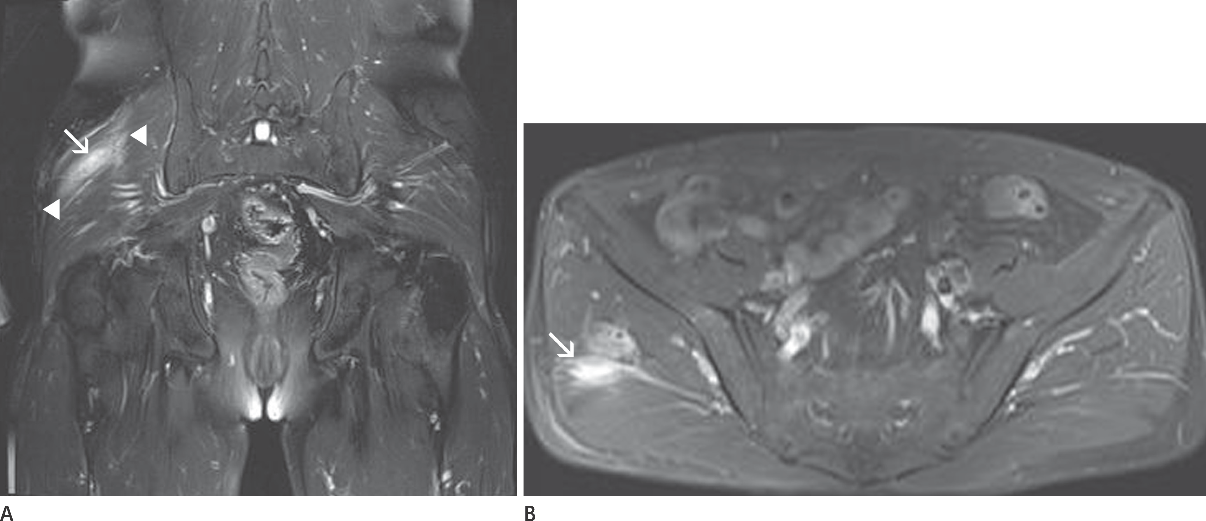
 | Fig. 1.A 50-year-old male patient diagnosed with oropharyngeal cancer two years previously and presently complaining of buttock pain. A. On coronal fat-suppressed T2-weighted image of the pelvis, a hyperintense soft tissue lesion with ill-defined margin (arrow) is seen in the muscular layer of the right buttock. Peritumoral edema (arrowheads) is also noted. B. The lesion shows homogeneous enhancement without degenerative change within the lesion (arrow) on axial fat-suppressed, contrast-enhanced T1-weighted image. |
 | Fig. 2.A 47-year-old male patient with multiple bone and soft tissue lesions. After biopsy for soft tissue lesion, lung cancer was diagnosed. A. On coronal fat-suppressed T2-weighted image of pelvis, two heterogeneous soft tissue masses with peritumoral edema are seen within each iliopsoas (arrow) and adductor muscle (arrowhead). B. On another scan of same sequence, multiple bone metastatic lesions are seen (arrows). C. Metastatic mass located within the right iliopsoas muscle shows degenerative change (arrow) on axial fat-suppressed contrast enhanced T1-weighted image. |
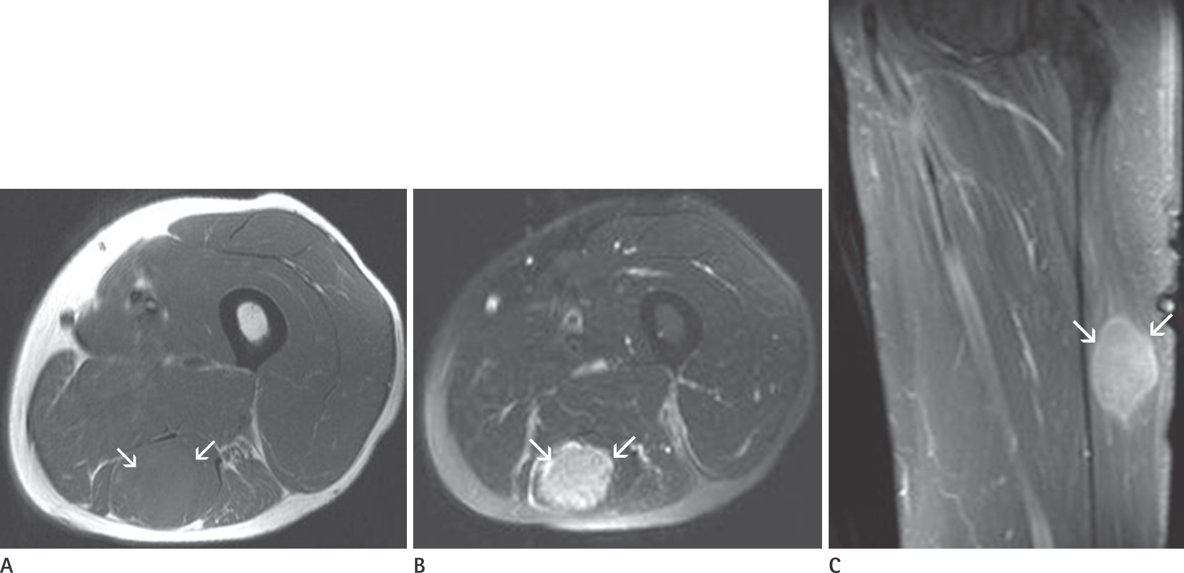
 | Fig. 3.A 67-year-old male patient with intramuscular metastasis and unknown lung cancer. A. On axial T1-weighted magnetic resonance image of left thigh, a homogeneous mass lesion (arrows) is seen in the semitendinosus muscle. B. The soft tissue lesion (arrows) shows well-defined margin without peritumoral edema on axial fat-suppressed T2-weighted image. C. On sagittal fat-suppressed, contrast-enhanced T1-weighted image, the mass lesion (arrows) shows homogeneous enhancement without degenerative change. |
Table 1.
The Types of Primary Malignancies and the Number of Patients Diagnosed with Soft Tissue Metastasis According to Each Primary Malignancy
Table 2.
The Anatomical Locations of Soft Tissue Lesions
Table 3.
Clinical and Magnetic Resonance Imaging Analysis Results for Differentiating between Soft Tissue Metastasis and Benign Soft Tissue Lesion


 PDF
PDF ePub
ePub Citation
 Print
Print


 XML Download