

 PDF
PDF ePub
ePub Citation
 Print
Print


INTRODUCTION
Cavernous hemangiomas are benign tumors rather than vascular malformations. They can occur in any internal organ, but are relatively rare in the intracranial extracerebral area (1). Despite a rare frequency, it is crucial to accurately interpret the imaging features because a wrong procedure based on misdiagnosis, caused by atypical imaging findings, can be fatal to patients, due to massive bleeding (2). In this context, we present the case of a 67-year-old woman with atypical imaging findings of a cavernous sinus cavernous hemangioma.
CASE REPORT
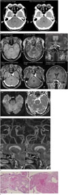
A 67-year-old woman visited the emergency room in our hospital with sudden onset of a persistent severe headache, with occasional dizziness and vomiting. Her mental status was alert, but initial systolic blood pressure was as high as 180 mm Hg. She had no specific past medical history. Contrast-enhanced computed tomography (CT) was performed to differentiate emergent conditions such as acute hemorrhage from non-emergent conditions. On CT images (Fig. 1A), a well-defined contour-bulge of about 24 × 18 × 14 mm was seen in the right cavernous sinus and right pituitary gland without definite evidence of hemorrhage. The mass showed high attenuation on pre-contrast enhancement images, with scattered high attenuation foci, suggesting tiny calcifications. On post-contrast enhancement images, the mass showed strong peripheral enhancement. Compared to the peripheral portion, the central portion showed poor enhancement. For further evaluation, contrast-enhanced magnetic resonance imaging (MRI) was performed (Fig. 1B). The mass showed heterogeneous slightly low signal intensity on the T1-weighted images (T1WI). On the T2-weighted images (T2WI), the mass showed overall high signal intensity, with a few small internal dark portions. Contrast enhanced images were performed with two-dimensional axial T1WI, immediately after intravenous administration of gadolinium contrast media. Delayed images were performed with three-dimensional (3D) T1 gradient echo. As in the contrast-enhanced CT images, on contrast-enhanced MRI, the mass showed peripheral enhancement, with a poorly enhanced central portion. The central portion showed no filling-in or delayed enhancement pattern. The posterior portion of the mass showed slightly high signal intensity on diffusion-weighted MRI and low signal intensity on the apparent diffusion coefficient map, meaning diffusion restriction, probably because of thrombosis or subacute hematoma (Fig. 1C). The right internal carotid artery passed through the mass, without luminal narrowing or obstruction (Fig. 1D). Furthermore, the pituitary stalk deviated to the left, on the opposite side of the mass, by the mass effect (Fig. 1B, thick arrow).
Based on these radiologic findings, we suspected a soft tissue mass with internal hemorrhage, such as an invasive pituitary adenoma. After 3 days, tumor removal was performed by a trans-sphenoidal approach. In the operating room, the neurosurgeon reported that lobulated-shaped dural-based mass such as meningioma was located in cavernous sinus. Microscopically, this mass was composed of various-sized dilated vascular spaces, lined by non-atypical endothelial cells. Fibrous tissue was present between the vascular spaces. Also, a blood clot, consisting of platelets, fibrin, and red blood cells was found in the intravascular space. In conclusion, this mass was pathologically confirmed as a cavernous hemangioma with internal thrombosis (Fig. 1E). The poorly enhancing areas in the mass, observed on imaging evaluations, correlated with an internal thrombus on microscopic evaluation.
DISCUSSION
Cavernous hemangioma is a benign tumor composed of vascular structures with proliferative endothelium and connective tissue and is most commonly located in the middle cranial fossa (3) within the cranium. It is essential to correctly comprehend the imaging findings of intracranial extracerebral cavernous hemangiomas because a wrong procedure based on misdiagnosis is fatal to the patient with 12.5–25% mortality rate (2). Additionally, the reported rate of misdiagnosis is as high as 38.9% (4).
In this case, a 67-year-old female patient visited the emergency room in our hospital because of a continuous severe headache and intermittent dizziness. These symptoms occurred suddenly on the hospital visitation date. Focal contour bulging mass with high attenuation and tiny calcifications on CT images and high signal intensity with internal dark foci on T2WI was detected. This mass had a peripheral enhancement, with a poorly enhanced central portion. After tumor removal, dark foci on T2WI and poorly central enhanced portion correlated with an internal thrombus on microscopic evaluation.
Unlike our case, according to the literature, typical symptoms are insidious, and headache is the most common symptom. Additionally, seizures, hemorrhages, and dysfunction of the cranial nerves in the cavernous sinus can also occur (5). The cause of these symptoms is not well-understood, but the hypothesis with the highest probability is that they are caused by progressive dilatation of the vascular component (5).
Known typical imaging findings of intracranial extracerebral cavernous hemangiomas are a well-defined contour-bulging mass, with marked homogeneous enhancement or a filling-in enhancement pattern, on post-contrast CT and MR images. Probably, this enhancement pattern could be because cavernous hemangiomas have a high vascularity with a variable circulation velocity, due to the feeding artery and numerous vascular channels of variable size (1, 6). Also, this mass shows intermediate-to-high attenuation on pre-contrast CT images and can be combined with calcifications. MRI shows homogeneously high signal intensity on proton density images and T2WI, and low signal intensity on T1WI (124).
The intracranial extracerebral cavernous hemangioma, in this case, was different from the typical imaging appearance. The most obvious difference was the internal irregular-shaped area, with poor enhancement and low signal intensity on T2WI. When correlated with the pathology, these imaging findings are caused by an internal thrombosis. On retrospection, we should have thought that the internal area with poor enhancement can be calcification or thrombosis or hematoma, rather than necrosis or a cystic change because of the dark signal on T2WI. Also, because this area is different from the calcification on pre-enhancement CT, it is most likely to be a thrombosis or hematoma.
According to the literature, besides the case presented in this report, intracranial extracerebral cavernous hemangioma can show various atypical imaging findings including mild-tomarked enhancement and various internal signal intensity, depending on whether hyaline degeneration, thrombosis, hematoma or deposition of minerals or collagen, has occurred. A ring- or tubular-shaped enhancement is also possible (5).
In conclusion, it is necessary to differentiate atypical cavernous hemangioma when there is evidence of hemorrhage or thrombosis in the parasellar mass invading the cavernous sinus.
 XML Download