

 PDF
PDF ePub
ePub Citation
 Print
Print


Abstract
Purpose
To determine adequate timing of acquisition of contrast-enhanced fluid attenuated inversion recovery (FLAIR) by using multiphasic contrast-enhanced FLAIR magnetic resonance imaging (MRI) and to evaluate added value in detecting small intracerebral metastases 5 mm or less.
Materials and Methods
Twenty-nine patients, that underwent multiphasic contrast-enhanced FLAIR MRI and contrast-enhanced T1 weighted image (T1WI) were included and total number of small intracerebral metastases was 131. Sensitivity, specificity and accuracy of lesion detection were evaluated. Contrast ratio (CR) and enhancement ratio of each lesion were compared and analyzed among each imaging sequence.
Results
Sensitivity, specificity and accuracy of lesion detection were increased when contrast-enhanced FLAIR was added to contrast-enhanced T1WI. Area of under receiver operating characteristic curve significantly increased by addition of contrast-enhanced FLAIR than using contrast-enhanced T1WI alone (p < 0.05). CR was significantly higher in contrast-enhanced T1WI than FLAIR (p < 0.001). All of the above results were not different according to time of acquisition of contrast-enhanced FLAIR.
Go to : 
References
1. Barnholtz-Sloan JS, Sloan AE, Davis FG, Vigneau FD, Lai P, Sawaya RE. Incidence proportions of brain metastases in patients diagnosed (1973 to 2001) in the Metropolitan Detroit Cancer Surveillance System. J Clin Oncol. 2004; 22:2865–2872.

2. Patchell RA. The management of brain metastases. Cancer Treat Rev. 2003; 29:533–540.
3. Ranjan T, Abrey LE. Current management of metastatic brain disease. Neurotherapeutics. 2009; 6:598–603.
4. Hall WA, Djalilian HR, Nussbaum ES, Cho KH. Long-term survival with metastatic cancer to the brain. Med Oncol. 2000; 17:279–286.
5. Stelzer KJ. Epidemiology and prognosis of brain metastases. Surg Neurol Int. 2013; 4(Suppl 4):S192–S202.
6. Chang EL, Hassenbusch SJ 3rd, Shiu AS, Lang FF, Allen PK, Sawaya R, et al. The role of tumor size in the radiosurgical management of patients with ambiguous brain metastases. Neurosurgery. 2003; 53:272–280. ; discussion 280–281.
7. Lin J, Jandial R, Nesbit A, Badie B, Chen M. Current and emerging treatments for brain metastases. Oncology (Williston Park). 2015; 29:250–257.
8. Ahn SJ, Chung TS, Chang JH, Lee SK. The added value of double dose gadolinium enhanced 3D T2 fluid-attenuated inversion recovery for evaluating small brain metastases. Yonsei Med J. 2014; 55:1231–1237.
9. Ercan N, Gultekin S, Celik H, Tali TE, Oner YA, Erbas G. Diagnostic value of contrast-enhanced fluid-attenuated inversion recovery MR imaging of intracranial metastases. AJNR Am J Neuroradiol. 2004; 25:761–765.
10. Fukuoka H, Hirai T, Okuda T, Shigematsu Y, Sasao A, Kimura E, et al. Comparison of the added value of contrast-enhanced 3D fluid-attenuated inversion recovery and magnetization-prepared rapid acquisition of gradient echo sequences in relation to conventional postcontrast T1-weighted images for the evaluation of leptomeningeal diseases at 3T. AJNR Am J Neuroradiol. 2010; 31:868–873.
11. Tomura N, Narita K, Takahashi S, Otani T, Sakuma I, Yasuda K, et al. Contrast-enhanced multi-shot echo-planar FLAIR in the depiction of metastatic tumors of the brain: comparison with contrast-enhanced spin-echo T1-weighted imaging. Acta Radiol. 2007; 48:1032–1037.
12. Lee EK, Lee EJ, Kim S, Lee YS. Importance of contrast-enhanced fluid-attenuated inversion recovery magnetic resonance imaging in various intracranial pathologic conditions. Korean J Radiol. 2016; 17:127–141.
13. Terae S, Yoshida D, Kudo K, Tha KK, Fujino M, Miyasaka K. Contrast-enhanced FLAIR imaging in combination with pre-and postcontrast magnetization transfer T1-weighted imaging: usefulness in the evaluation of brain metastases. J Magn Reson Imaging. 2007; 25:479–487.
14. Essig M, Knopp MV, Schoenberg SO, Hawighorst H, Wenz F, Debus J, et al. Cerebral gliomas and metastases: assessment with contrast-enhanced fast fluid-attenuated inversion-recovery MR imaging. Radiology. 1999; 210:551–557.
15. Mathews VP, Caldemeyer KS, Lowe MJ, Greenspan SL, Weber DM, Ulmer JL. Brain: gadolinium-enhanced fast fluid-attenuated inversion-recovery MR imaging. Radiology. 1999; 211:257–263.
16. Mathews VP, Caldemeyer KS, Ulmer JL, Nguyen H, Yuh WT. Effects of contrast dose, delayed imaging, and magnetization transfer saturation on gadolinium-enhanced MR imaging of brain lesions. J Magn Reson Imaging. 1997; 7:14–22.
17. Melhem ER, Bert RJ, Walker RE. Usefulness of optimized gadolinium-enhanced fast fluid-attenuated inversion recovery MR imaging in revealing lesions of the brain. AJR Am J Roentgenol. 1998; 171:803–807.
18. Yuh WT, Tali ET, Nguyen HD, Simonson TM, Mayr NA, Fisher DJ. The effect of contrast dose, imaging time, and lesion size in the MR detection of intracerebral metastasis. AJNR Am J Neuroradiol. 1995; 16:373–380.
19. Jeon JY, Choi JW, Roh HG, Moon WJ. Effect of imaging time in the magnetic resonance detection of intracerebral metastases using single dose gadobutrol. Korean J Radiol. 2014; 15:145–150.
20. Cohen-Inbar O, Xu Z, Dodson B, Rizvi T, Durst CR, Mukherjee S, et al. Time-delayed contrast-enhanced MRI improves detection of brain metastases: a prospective validation of diagnostic yield. J Neurooncol. 2016; 130:485–494.
21. Kushnirsky M, Nguyen V, Katz JS, Steinklein J, Rosen L, War-shall C, et al. Time-delayed contrast-enhanced MRI improves detection of brain metastases and apparent treatment volumes. J Neurosurg. 2016; 124:489–495.
22. Kremer S, Abu Eid M, Bierry G, Bogorin A, Koob M, Dietemann JL, et al. Accuracy of delayed post-contrast FLAIR MR imaging for the diagnosis of leptomeningeal infectious or tumoral diseases. J Neuroradiol. 2006; 33:285–291.
23. Hirota T, Ishihara K, Akazawa K, Kubota T, Yamada K, Nishimura T. Case report: delayed post-contrast fluid-attenuated inversion recovery image for depicting meningeal carcinomatosis. Br J Radiol. 2004; 77:528–531.
24. Kazi AZ, Joshi PC, Kelkar AB, Mahajan MS, Ghawate AS. MRI evaluation of pathologies affecting the corpus callosum: a pictorial essay. Indian J Radiol Imaging. 2013; 23:321–332.
25. Yoshida A, Tha KK, Fujima N, Zaitsu Y, Yoshida D, Tsukahara A, et al. Detection of brain metastases by 3-dimensional magnetic resonance imaging at 3 T: comparison between T1-weighted volume isotropic turbo spin echo acquisition and 3-dimensional T1-weighted fluid-attenuated inversion recovery imaging. J Comput Assist Tomogr. 2013; 37:84–90.
26. Kato Y, Higano S, Tamura H, Mugikura S, Umetsu A, Murata T, et al. Usefulness of contrast-enhanced T1-weighted sampling perfection with application-optimized contrasts by using different flip angle evolutions in detection of small brain metastasis at 3T MR imaging: comparison with magnetization-prepared rapid acquisition of gradient echo imaging. AJNR Am J Neuroradiol. 2009; 30:923–929.
Go to :
 | Fig. 1.53-year-old female with brain metastases from non-small cell lung cancer. On axial image of postcontrast T1 weighted image (A), there is a suspicious enhancing nodular lesion in left frontal area confused with cortical vessels (arrow). And axial image of precontrast fluid attenuated inversion recovery (B) shows perilesional edema around the lesion (arrow). Therefore the lesion was assessed as metastasis, not a cortical vessel. |
 | Fig. 2.57-year-old male with brain metastases from non-small cell lung cancer. Axial image of postcontrast T1 weighted image (A) shows suspicious enhancing nodular lesion in left cerebellar hemisphere (arrow). But axial images of precontrast FLAIR (B), 1st postcontrast FLAIR (C), and 2nd postcontrast FLAIR (D) do not show abnormal enhancement at the same location. Therefore the lesion was assessed as pulsating artifact, not a metastasis. FLAIR = fluid attenuated inversion recovery |
 | Fig. 3.78-year-old male with brain metastases from colon cancer. Axial image of postcontrast T1 weighted image (A) shows a subtle enhancing nodule in right frontal cortex (arrow). And axial images of 1st postcontrast FLAIR (B) and 2nd postcontrast FLAIR (C) also show strong rim-enhancing nodule at the same location (arrows). Therefore the lesion was assessed as metastasis. FLAIR = fluid attenuated inversion recovery |
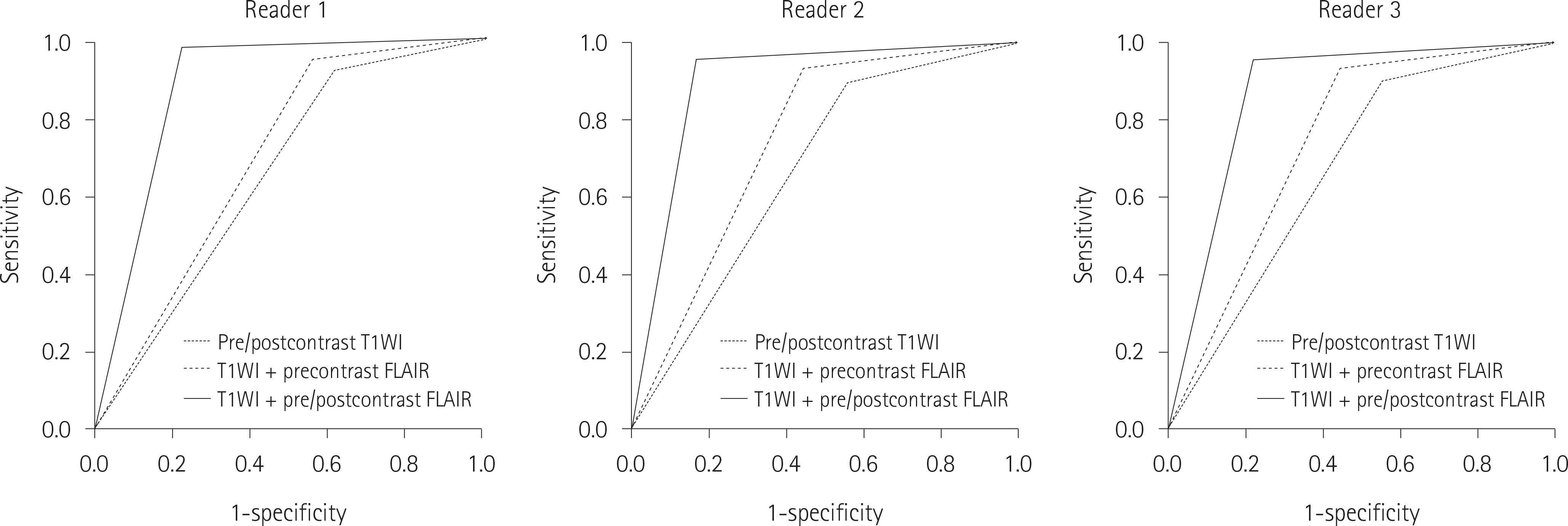 | Fig. 4.ROC curves for detectability of small brain metastases of three readers. The area of under ROC curve is significantly greater in T1WI + pre/ postcontrast FLAIR as compared to that in pre/postcontrast T1WI or T1WI + precontrast FLAIR. FLAIR = fluid attenuated inversion recovery, ROC = receiver operating characteristic, T1WI = T1 weighted image |
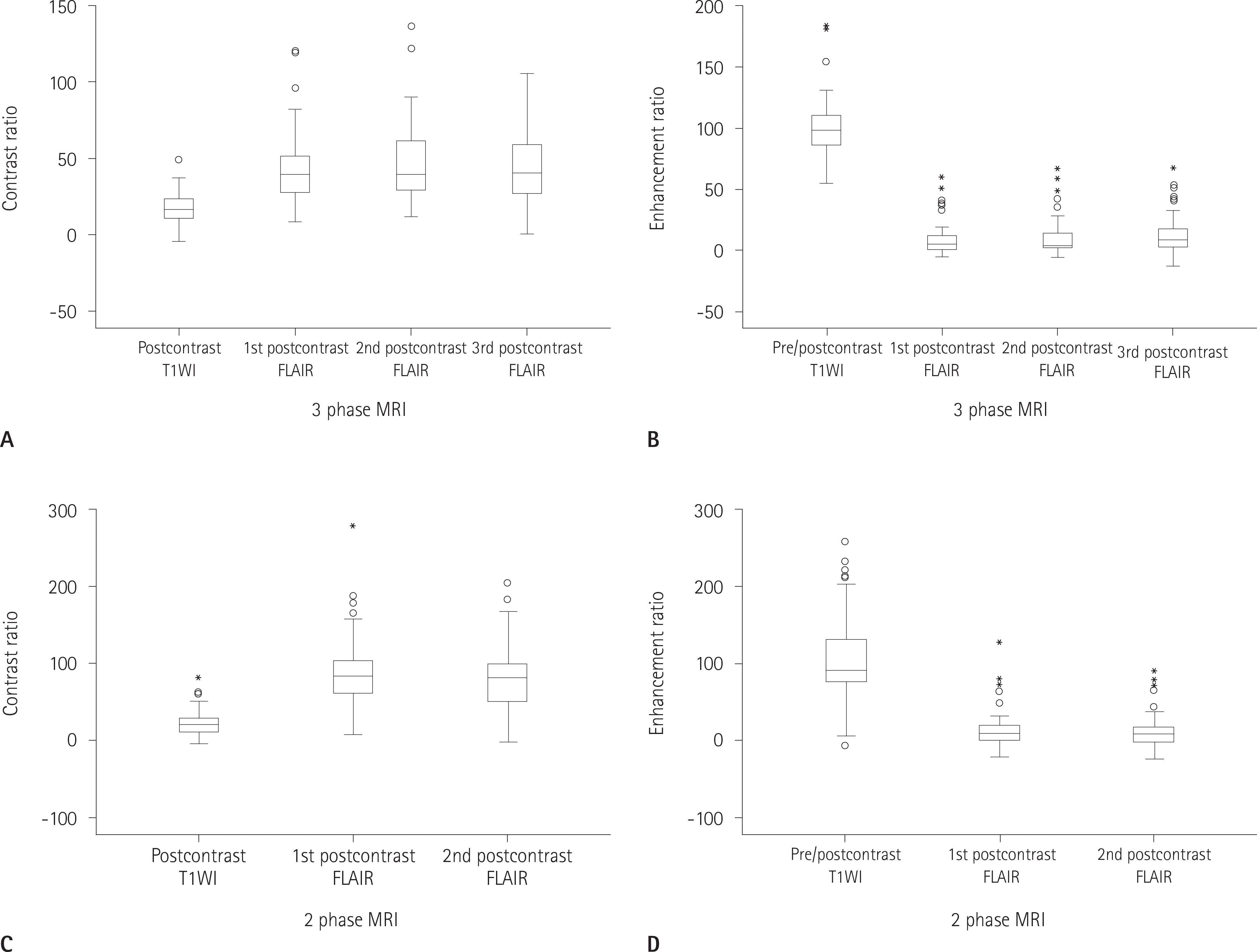 | Fig. 5.Comparison of CRs and ERs on multiphase contrast enhanced FLAIR MRI. In 3-phase MRI (A, B), CRs of small brain metastases in post-contrast T1WI are significantly lower than those in postcontrast FLAIR (p < 0.001) and ERs in pre/postcontrast T1WI are significantly greater than those in pre/postcontrast FLAIR (p < 0.001). In 2-phase MRI (C, D), the result is not different from the above. And there is no significant difference between CRs and ERs with time delays in both 2-phase and 3-phase contrast enhanced FLAIR MRI. CR = contrast ratio, ER = enhancement ratio, FLAIR = fluid attenuated inversion recovery, MRI = magnetic resonance imaging, T1WI = T1 weighted image |
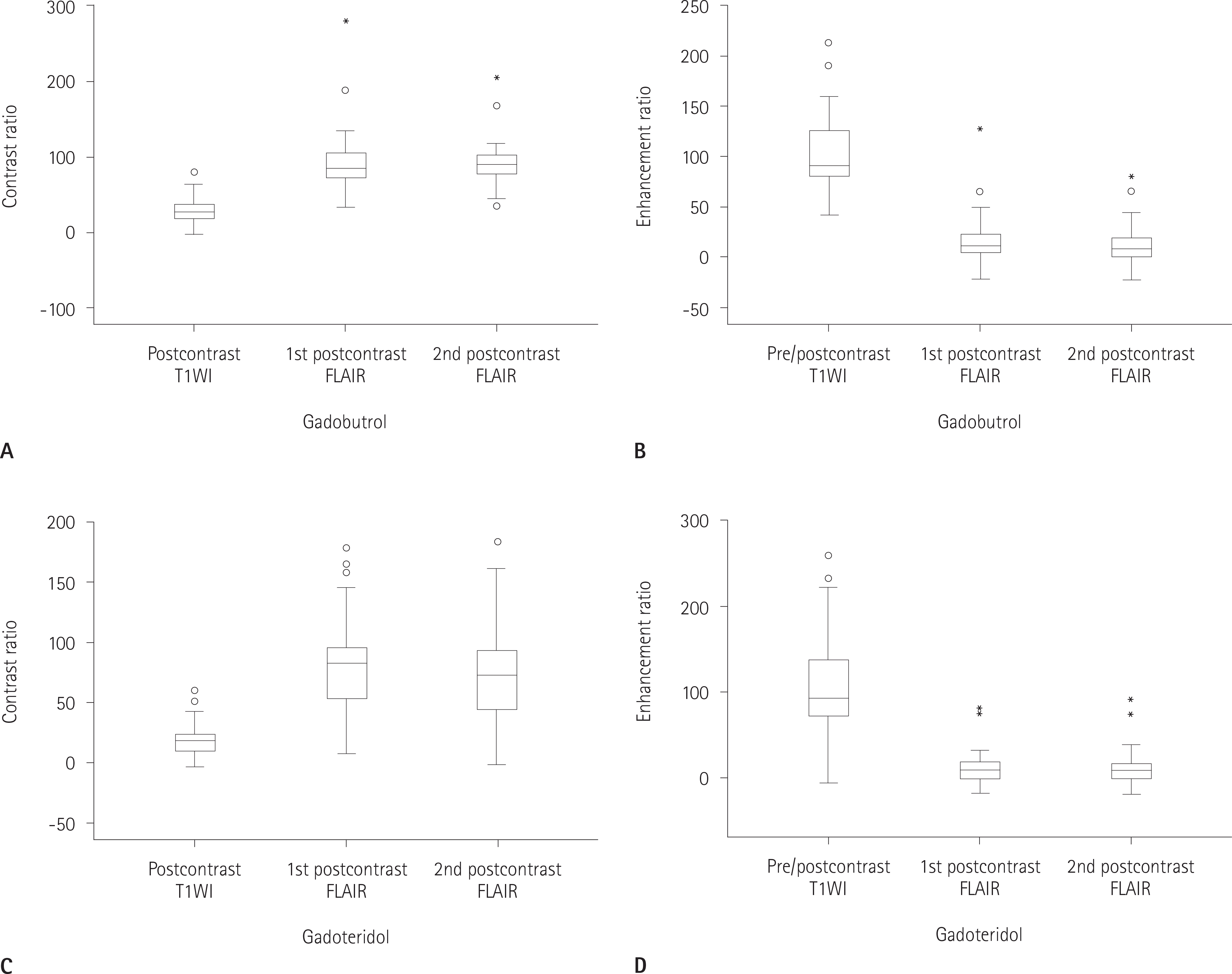 | Fig. 6.Comparison of CRs and ERs for contrast media on 2 phase contrast enhanced FLAIR magnetic resonance imaging. When gadobutrol is used (A, B), CRs of small brain metastases in postcontrast T1WI are significantly lower than those in postcontrast FLAIR (p < 0.001) and ERs in pre/postcontrast T1WI are significantly greater than those in pre/postcontrast FLAIR (p < 0.001). When gadoteridol is used (C, D), the result is not different from the above. When gadobutrol and gadoteridol are used, there is no difference in CRs and ERs according to time delays. CR = contrast ratio, ER = enhancement ratio, FLAIR = fluid attenuated inversion recovery, T1WI = T1 weighted image |
Table 1.
Sensitivity, Specificity, and Diagnostic Accuracy in Three Readers
Because the results of 1st, 2nd, and 3rd postcontrast FLAIR are the same, the results of 2nd and 3rd postcontrast FLAIR are not mentioned in the table. Post FLAIR = 1st (or 2nd or 3rd) postcontrast fluid attenuated inversion recovery, Post T1WI = postcontrast T1 weighted image, Pre FLAIR = precontrast fluid attenuated inversion recovery, Pre T1WI = precontrast T1 weighted image
Table 2.
Area of Under Receiver Operating Characteristic Curve
Table 3.
Effect of Time Delay after Contrast Injection on Multiphase CE-FLAIR
CE-FLAIR = contrast enhanced fluid attenuated inversion recovery, CR = contrast ratio, ER = enhancement ratio, SD = standard deviation, T1WI = T1 weighted image, 1 = precontrast T1WI + postcontrast T1WI, 2 = precontrast FLAIR + 1st postcontrast FLAIR, 3 = precontrast FLAIR + 2nd postcontrast FLAIR, 4 = precontrast FLAIR + 3rd postcontrast FLAIR
Table 4.
Effect of Time Delay after Gadobutrol or Gadoteridol Injection on 2 Phase Contrast Enhanced FLAIR
 XML Download