

 PDF
PDF ePub
ePub Citation
 Print
Print


INTRODUCTION
Intraneural hemangioma is quite rare and occurs most frequently in the median nerve (12345). Only a few cases of intraneural hemangiomas of the median nerve have been described in the literature (123). Recently, we experienced a case of 38-year-old-man with intraneural hemangioma of the median nerve. The patient had a small palpable mass in the volar aspect of the wrist and symptoms of carpal tunnel syndrome. And he had no history of recent trauma. Initially, the mass was misdiagnosed as an in-continuity neuroma, due to lack of significant blood flow on color Doppler imaging and uncertain continuity of individual nerve fascicle. We report ultrasound (US) and magnetic resonance imaging (MRI) findings of a case with intraneural hemangioma of the median nerve with literature review.
CASE REPORT
A 38-year-old-man presented with pain and tingling sensation in the right hand for several months and his symptoms worsened for a few days. Tingling sensation was noted in the thumb, index, middle and radial half of the ring fingers. There was no motor weakness in the hand. He has participated in computer-related work and has been used the keyboard and the computer mouse.
There was no history of trauma and relevant medical condition. Physical examination revealed a small palpable mass in the volar aspect of the wrist, just proximal to the flexor retinaculum. There was no tenderness, but Tinel's sign and Phalen's sign were positive. Under the impression of carpal tunnel syndrome, US was performed initially to determine the nature of the mass and the status of the median nerve.
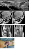
US of the median nerve, just proximal to the flexor retinaculum, revealed a swollen nerve containing a 1.9 × 0.9 cm-sized, infiltrative hypoechoic mass separating the nerve fascicles (Fig. 1A, long axis). Color Doppler imaging with compression test showed no significant blood flow within the mass (Fig. 1A, short axis). Continuity of each individual nerve fascicle was difficult to identify, except the peripherally displaced fascicles. Our initial differential diagnosis included lipomatosis of nerve, in-continuity neuroma, and intraneural hemangioma, based on the intraneural location and infiltrative pattern of the mass. Because we could not verify the continuity of individual nerve fascicle and the mass showed no significant blood flow, in-continuity neuroma was considered as first. The median nerve within the carpal tunnel shows no abnormality.
MRI was performed for better characterization of the mass, and showed an infiltrative intraneural mass in the median nerve just proximal to the flexor retinaculum. The mass showed heterogeneous but predominantly high signal intensity on T2-weighted image (Fig. 1B, D) and low signal intensity containing central hyperintense area on T1-weighted image (Fig. 1C). The nerve fascicles were separated and displaced peripherally by the intraneural mass. Central hyperintense area on T1-weighted image was considered to indicate hemorrhagic component, but this component could not verify on the retrospective assessment of the US images. The mass showed well heterogeneous enhancement on the contrast-enhanced fat-saturated T1-weighted image (Fig. 1E).
The operation was performed under general anesthesia, using a pneumatic tourniquet. Microsurgical interfascicular dissection of the median nerve and resection of the entire tumor were performed, without damaging the nerve fibers. Intraoperative findings showed a blue mass with about 2 cm-sized of median nerve (Fig. 1F). The histopathologic examination revealed irregular shaped and dilated vascular structure in the loose fibrous stroma and confirmed the diagnosis of a capillary hemangioma arising from the median nerve. There was no recurrence of symptoms during the follow-up period of 1 year.
DISCUSSION
Intraneural hemangiomas of peripheral nerves are extremely rare. In the literature, intraneural hemangiomas were reported in median nerve, posterior tibial nerve, ulnar nerve, and digital nerves, and have a predilection for the median nerve (123456). About fifteen cases of intraneural hemangiomas of the median nerve have been reported. A persistent median artery has been advocated to explain the prevalence of the hemangioma in the median nerve (2).
Peripheral nerve tumors comprise less than 5% of all tumor of the hand, which can be divided into neural and non-neural origin. Intraneural tumors of neural origin composed of schwannoma and neurofibroma. Nerve tumors, which are intraneural in location but non-neural in origin, include lipomatosis of nerve, intraneural lipomas, hemangiomas, and ganglion cysts (4). In our case, initial differential diagnosis include lipomatosis of nerve, traumatic neuroma, and intraneural hemangioma, based on the intraneural location and infiltrative pattern on US and patient's presenting symptoms.
Lipomatosis of nerve refers to a tumor-like nerve disorder with epineural and perineural infiltration by adipose and fibrous tissue (7). US findings with infiltrative mass separating the nerve fascicles in our case warranted consideration of a lipomatosis of the median nerve. But neither US nor MRI revealed typical coaxial cable-like appearance, and because of the lack of fat signal on the MRI, the diagnosis of lipomatosis was excluded.
Traumatic neuromas can be classified into amputation neuroma and in-continuity neuroma. The amputation neuroma is localized at the proximal stump of the nerve and the in-continuity neuroma connects the two nerve stumps with variable saving of nervous fascicles (8). Most traumatic neuromas show intermediate signal intensity on T1-weighted image and inhomogeneous intermediate to high signal intensity on T2-weighted image. On the US, these masses are often hypoechoic but heterogeneous, and continuity between the mass and a nerve is essential to the diagnosis. US findings of our case were a hypoechoic infiltrative mass with lack of blood flow and uncertain continuity of individual nerve fascicle, and so, in-continuity neuroma was the first consideration. But our patient had no traumatic or surgical history, and the mass showed heterogeneous but predominantly high signal intensity on T2-weighted MR image.
US and MRI are widely regarded as the standard imaging modality for evaluating hemangiomas (9). On the US, hemangiomas may have a heterogeneous appearance, with a variable echogenicity, ranging from hypoechoic to hyperechoic relative to surrounding soft tissue, which often infiltrates the involved soft tissue. Color Doppler imaging shows increased blood flow, but may detect only sparse flow in the slow-flow or involuted hemangioma, in which cases compression test may show blood flow inside a lesion. Hemangiomas usually show intermediate to decreased signal intensity on T1-weighted images and increased signal intensity on T2-weighted and STIR images. In cases of thrombosis or hemorrhage, heterogeneous signal intensity can be observed (10). The lesion in our case was seen as an infiltrative mass with heterogeneous but predominantly high signal intensity on T2-weighted image and well heterogeneous enhancement, and these MRI findings were helpful for correct diagnosis on the retrospective review. We initially misdiagnosed as an in-continuity neuroma because authors were less familiar with the intraneural hemangioma and US showed no significant blood. Lack of blood flow may be related to thrombosis or hemorrhage within the lesion.
The most described treatment for intraneural hemangioma is interfascicular dissection and complete excision of tumor if possible, and it is important to avoid damage to the nerve structure (5). But it is not always possible to completely remove the intraneural hemangioma safely without damage to the nerve fascicles, and if partially removed from the median nerve, there is a high risk of recurrence. So, correct preoperative diagnosis is important for operative planning and informed consent of the patients.
Intraneural hemangioma is extremely rare and is not familiar to radiologists. Here, we report a case of intraneural hemangioma of median nerve, which was seen as an infiltrative intraneural mass and showed heterogeneous but predominantly high signal intensity on T2-weighted image and well heterogeneous enhancement. We initially misdiagnosed as an in-continuity neuroma due to lack of blood flow within the mass, but awareness of imaging findings of intraneural hemangioma may guide to a correct diagnosis.
 XML Download