

 PDF
PDF ePub
ePub Citation
 Print
Print


Abstract
Purpose
To evaluate the magnetic resonance imaging (MRI) findings to improve the diagnostic accuracy for transverse process fractures and sacral fractures.
Materials and Methods
The lumbosacral MRI scans of 214 patients (mean age, 60 years; male-to-female ratio, 85:129), who had spine trauma between January and November 2015 were included. Two radiologists evaluated the presence, number, level, and anatomic site of the fractures on MRI with computed tomography as reference standard. Imaging findings were described as cortical disruption, marrow edema, or soft tissue edema on T1-, T2-, and fat-suppressed T2-weighted images. A statistical analysis was performed to compare the diagnostic accuracy of the MRI pulse sequences for the transverse process and sacral fractures.
Results
Of 168 fractures, 26 (15.5%) and 13 (4.9%) were in the transverse processes and sacra, respectively. A paravertebral soft tissue edema occurred in the transverse process fractures (80.8%) and presacral soft tissue and marrow edemas occurred in the sacral fractures (46.1%). The sensitivity for the transverse process fractures was 88% on the T2-weighted image. It was 92% on fat- suppressed T2-and T1-weighted images for sacral fractures.
Go to : 
References
1. Denis F. The three column spine and its significance in the classification of acute thoracolumbar spinal injuries. Spine (Phila Pa 1976). 1983; 8:817–831.

2. Sturm JT, Perry JF Jr. Injuries associated with fractures of the transverse processes of the thoracic and lumbar vertebrae. J Trauma. 1984; 24:597–599.
3. Patten RM, Gunberg SR, Brandenburger DK. Frequency and importance of transverse process fractures in the lumbar vertebrae at helical abdominal CT in patients with trauma. Radiology. 2000; 215:831–834.
4. Denis F, Davis S, Comfort T. Sacral fractures: an important problem. Retrospective analysis of 236 cases. Clin Orthop Relat Res. 1988; 227:67–81.
5. Pizones J, Sánchez-Mariscal F, Zúñiga L, Álvarez P, Izquierdo E. Prospective analysis of magnetic resonance imaging accuracy in diagnosing traumatic injuries of the posterior ligamentous complex of the thoracolumbar spine. Spine (Ph-ila Pa 1976). 2013; 38:745–751.
6. Speer KP, Spritzer CE, Bassett FH 3rd, Feagin JA Jr, Garrett WE Jr. Osseous injury associated with acute tears of the anterior cruciate ligament. Am J Sports Med. 1992; 20:382–389.
7. Niall DM, Bobic V. Bone bruising and bone marrow edema syndromes: incidental radiological findings or harbingers of future joint degeneration? International Society of Arthroscopy, Knee Surgery and Orthopaedic Sports Medicine (ISAKOS). Available at:. http://www.isakos.com/innovations/niall.aspx. Accessed Jan 20,. 2012.
8. Mink JH, Deutsch AL. Occult cartilage and bone injuries of the knee: detection, classification, and assessment with MR imaging. Radiology. 1989; 170(3 Pt 1):823–829.
9. Sadineni RT, Pasumarthy A, Bellapa NC, Velicheti S. Imaging patterns in MRI in recent bone injuries following negative or inconclusive plain radiographs. J Clin Diagn Res. 2015; 9:TC10–TC13.
10. Krueger MA, Green DA, Hoyt D, Garfin SR. Overlooked spine injuries associated with lumbar transverse process fractures. Clin Orthop Relat Res. 1996; 327:191–195.
11. Berry GE, Adams S, Harris MB, Boles CA, McKernan MG, Collinson F, et al. Are plain radiographs of the spine necessary during evaluation after blunt trauma? Accuracy of screening torso computed tomography in thoracic/lumbar spine fracture diagnosis. J Trauma. 2005; 59:1410–1413. ;discussion 1413.
12. Cabarrus MC, Ambekar A, Lu Y, Link TM. MRI and CT of insufficiency fractures of the pelvis and the proximal femur. AJR Am J Roentgenol. 2008; 191:995–1001.
13. McNally EG, Wilson DJ, Ostlere SJ. Limited magnetic resonance imaging in low back pain instead of plain radiographs: experience with first 1000 cases. Clin Radiol. 2001; 56:922–925.
14. Wang B, Fintelmann FJ, Kamath RS, Kattapuram SV, Rosenthal DI. Limited magnetic resonance imaging of the lumbar spine has high sensitivity for detection of acute fractures, infection, and malignancy. Skeletal Radiol. 2016; 45:1687–1693.
Go to :
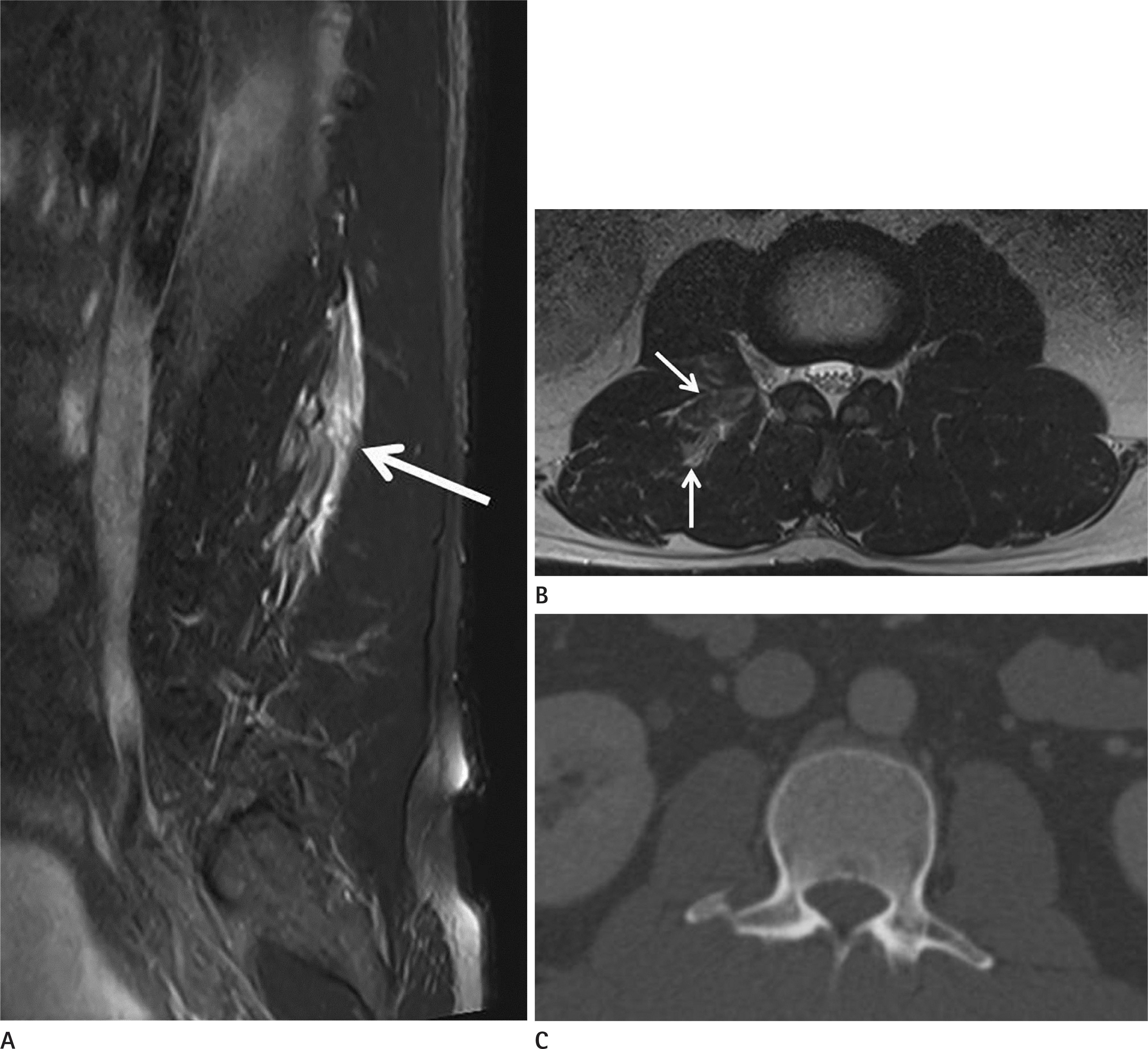 | Fig. 1.A 45-year-old man who incurred right L2 and L3 transverse process fractures after falling from a 2m high guardrail. A. Sagittal fat-suppressed T2-weighted MR image showing paravertebral soft tissue edema (arrow). B. Axial T2-weighted MR image showing soft tissue edema in the right psoas and spinal erector muscles (arrows), which reflects a transverse process fracture. C. Axial computed tomography scan showing a fracture at the right L3 transverse process. MR = magnetic resonance |
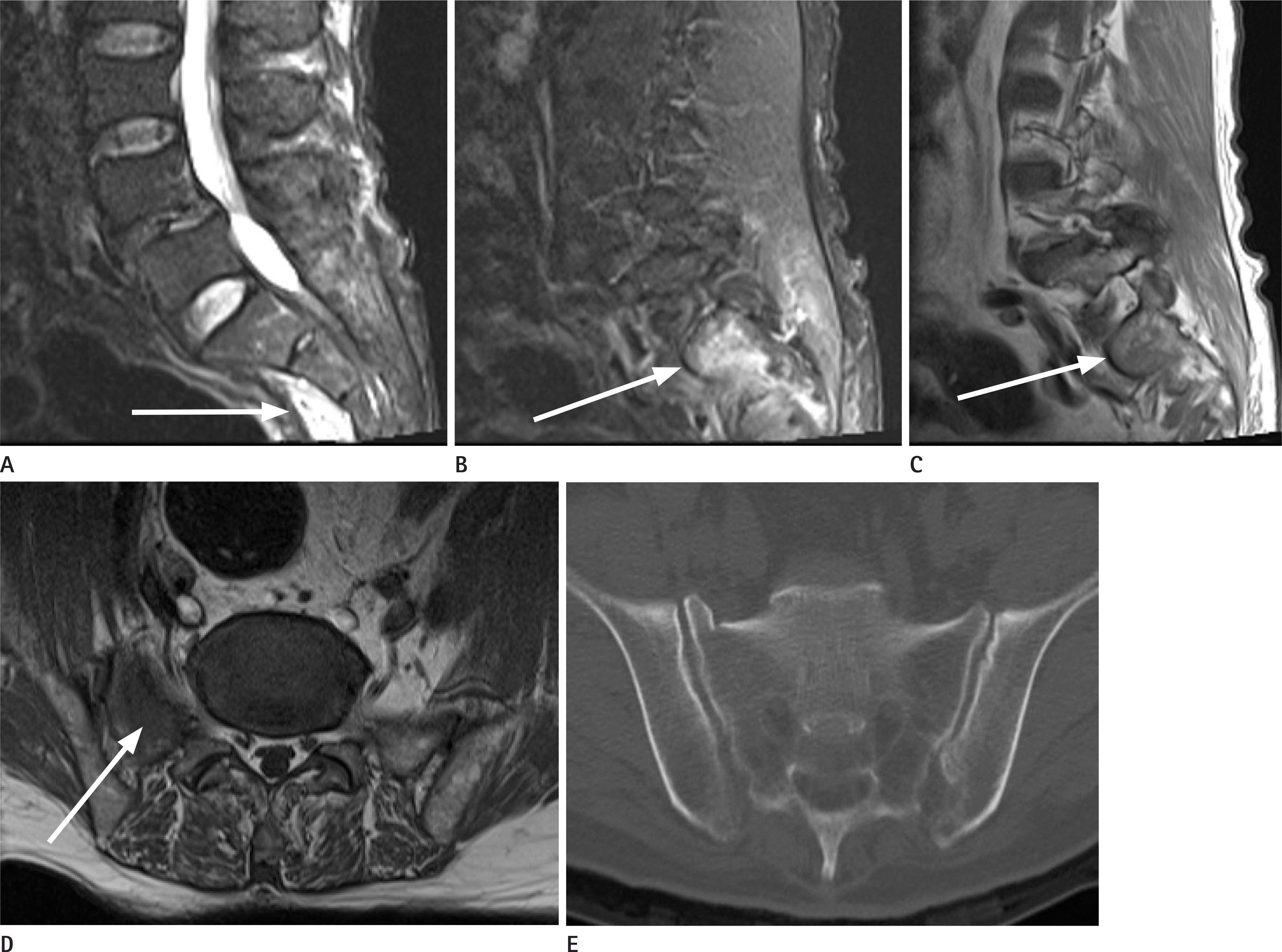 | Fig. 2.A 67-year-old man who incurred a trauma with a sacral fracture during a car accident. A. Midline sagittal fat-suppressed T2-weighted image showing presacral edema (arrow). B, C. Parasagittal fat-suppressed T2-weighted (B) and T1-weighted (C) images showing bone marrow edema (arrows) in the right sacral ala. D. Axial T2-weighted image showing bone marrow edema (arrow) in right sacral ala. E. Axial CT scan showing a fracture at the right sacral ala. |
 | Fig. 3.A false-positive case: a 78-year-old woman who had a low back pain after slipping down. A, B. Sagittal fat-suppressed T2-weighted (A) and T1-weighted (B) images showing presacral edema (arrows). C. No fracture can be observed in the sacrum on the sagittal computed tomography scan. |
Table 1.
The Diagnostic Accuracy of Magnetic Resonance Imaging in Spinal Fractures According to Fracture Sites and Pulse Sequences
 XML Download