

 PDF
PDF ePub
ePub Citation
 Print
Print


Dear Editor:
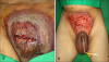
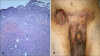
Pyoderma gangrenosum (PG) is a rare inflammatory ulcerative skin disease associated with systemic disease. Several cases have shown a relationship between PG and hematologic malignancies such as myelodysplastic syndrome (MDS). A 43-year-old man visited the department of plastic surgery because of a painful skin lesion on his suprapubic area since 1 month. He was highly obese (height, 177 cm; weight, 130 kg) and diagnosed as having diabetes mellitus 5 years ago. Physical examination revealed a solitary, large, erythematous round plaque, 15×5 cm in size, with a raised border and surrounding violaceous patches (Fig. 1). Initial laboratory data revealed normal white blood cell (WBC) count (5,680/µl), anemia (red blood cell [RBC] count, 2.3×10⁶/µl; hemoglobin level, 8.5 g/dl; hematocrit concentration, 25%), and low platelet count (84,000/µl). In the plastic surgery department, wound debridement, split-thickness skin graft placement, and surgical biopsy were performed. Biopsy revealed diffuse infiltration of neutrophils and lymphocytes in the epidermis, dermis, and subcutaneous fat tissue. Clinical and histopathological findings indicated PG (Fig. 2A). After debridement, the wound had become larger than initial size. He was referred to the dermatological department for cotreatment with methylprednisolone (125 mg/d) and cyclosporine (300 mg/d). During the treatment, he showed marked hematologic abnormalities and aggravated pancytopenia (WBC count, 1,800/µl; RBC count, 2.63×10⁶/µl; hemoglobin level, 8.5 g/dl; and platelet count, 31,000/µl). Thus, bone marrow biopsy was performed. The numbers of erythroid precursors and megakaryocytes increased, and granulocytic precursors relatively decreased. Accordingly, he was diagnosed as having MDS and referred to an oncologist for chemotherapy. For 5 months, he was treated with cyclosporine (300 mg/d) and methylprednisolone, with gradual tapering of the steroid dose (Fig. 2B). As a result, the PG lesion almost healed, but the MDS persisted. PG is a rare inflammatory, non-infectious, ulcerating skin disorder with an unknown pathogenesis, although immune complex-mediated neutrophil hyperactivity has been proposed123. Histopathological findings are not diagnostic but help to exclude other differential diagnoses. It usually shows neutrophilic and mixed lymphocytic infiltrations in the dermis1,3. No confirmatory diagnostic test is available, and diagnosis relies on clinical and histological correlation2. Immunosuppressive agents such as high-dose steroids, cyclosporine, and azathioprine can be used as treatment for PG. PG was frequently associated with systemic diseases such as inflammatory bowel disease, Crohn disease, Bechet disease, and hematologic malignancies12345. MDS is a hematologic malignancy that shows a neoplastic change in myeloid stem cells. It may also be associated with PG345. The pathogenesis of the association between PG and MDS is unknown. Some hypotheses have been proposed, including immunologic mediated processes involving the product of autoantibodies to cutaneous antigen and the deposition of immune complex. Patients with MDS have abnormal laboratory findings such as anemia, thrombocytopenia, and pancytopenia. Our patient showed aggravated abnormal laboratory findings during the diagnosis and treatment of PG. Based on the bone marrow biopsy result, we made a diagnosis of MDS. Dermatologists should diagnose PG expeditiously because of its rapid clinical course and association with other systemic diseases. Patients with PG should be evaluated for hematologic abnormalities. If abnormal findings are present, early bone marrow biopsy is needed to detect hematological malignancies.
 XML Download