

 PDF
PDF ePub
ePub Citation
 Print
Print


INTRODUCTION
Malignant melanoma (MM), either primary or metastatic can rarely mimics benign blue nevus (BN), some of which can cause diagnostic difficulties due to the absence of obvious cytologic atypia, typical of conventional MM123456.
Cytomorphologic features of melanoma cells of conventional type identified in the urine cytology specimen have been described in several reports. Usually, cytologic detection of melanoma cells of conventional melanoma is not problematic as they exhibit characteristic cytologic features including high nuclear to cytoplasmic ratio, macronucleoli and anisocytosis78. However, there have been no reports of blue nevus-like metastasis of urinary tract detected by urine cytology, which could be a diagnostic pitfall owing to their bland cytomorphology. Herein, we present an extremely rare case of blue nevus-like melanoma (BNLM) and its blue nevus like metastasis, the latter of which was identified in the catheterized urine cytology.
CASE REPORT
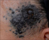
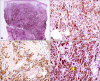
A 61-year-old man presented with blue-colored papuloplaques on his left temple that had been present for over 1 year. Over the previous 2 months, the lesions had grown rapidly, without any symptoms. Physical examination revealed a large ill-defined dark blue patch studded with pinhead to bean sized cobblestone surfaced papulonodules and satellite papules (Fig. 1). Punch biopsies were performed in the center and at the periphery of the plaque. Microscopic examination of the central specimen revealed compact sheet-like proliferation of heavily pigmented epithelioid and, occasionally, short spindle cells in the entire dermis and subcutaneous fat layer, without junctional activity. One focus of tumor necrosis was found. The tumor cells showed mild to moderate nuclear enlargement and inconspicuous nucleoli. The mitotic count was up to 4 per 10 high power fields with deep and mid-dermal mitotic cells. The Breslow thickness was 4.53 mm. In the periphery of the lesion, the overall pattern of tumor cell infiltrates was similar to that of the center. However, many of the tumor cells had small pyknotic nuclei and the lesser remainders showed mild to (rarely) moderate nuclear enlargement, vesicular nuclei, and small nucleoli. There were rare exceptions of overtly atypical cells showing hyperchromatic enlarged nuclei, and bizarre giant cells. Due to these atypical cells, albeit rare, the histologic findings of peripheral lesion are not compatible with those of other benign dermal melanocytic lesions, i.e., common or cellular blue nevus and Ota nevus. Furthermore, there was no necrosis or mitosis in the dermis (Fig. 2). The tumor cells of both lesions were positive to HMB45, Melan-A and S-100 protein immunohistochemical staining. MIB-1 labelling index was 22%. Considering the clinicohistopathological findings, we diagnosed both lesions as BNLM.
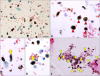
Following the punch biopsy, a fluorodeoxyglucose-positron emission tomography scan revealed multiple lymph node and bone metastases, but no remarkable findings in the urinary tract. The patient was admitted for chemotherapy with dacarbazine 407 mg/day (250 mg/m2 body surface area). During the admission, hematuria of unknown duration was detected. Catheterized urine cytology on SurePath liquid-based preparation was performed. This demonstrated many singly scattered, pigmented cells admixed with red blood cells and occasional necrotic cells. The cells were oval to spindle-shaped with blunt ends and showed a moderate degree of variation in shape and size. Although difficult to recognize nuclear details due to heavy melanin pigment in many of the cells, most cells had small nuclei; only a few cells had nuclei showing mild to moderate nuclear enlargement and nuclear hyperchromasia. Most of the cells on the smear were positive for HMB45 and Melan-A; hence, they were determined to be metastatic BN-like melanoma cells (Fig. 3). However, some histiocytes and urothelial cells were negative for both stains. Subsequent abdominopelvic computed tomography revealed possible liver, spleen, and right kidney metastases, and suggested potential metastases in the bladder dome and lungs. Owing to the patient's poor general condition, cystoscopy with biopsy was not performed. The patient died from pneumonia 3 months after the initial diagnosis.
DISCUSSION
Primary or metastatic MM can mimic benign BN in rare cases, making the diagnosis challenging123456. The former was known as so-called ‘malignant blue nevus (MBN)4.’ However, this term MBN can cause confusion not only as a nevus is, by definition, benign, but also as it can be applied to several different situation including 1) MM arising in association with a preexisting BN and 2) less likely melanoma with architectural or cytologic features resembling BN but arising de novo. Thus, it has been suggested that the former should be designated as “melanoma arising in association with BN” or “melanoma associated with BN,” and the latter as “blue nevus-like melanoma5.” Of these, the present case represents a BNLM. Diagnosing BNLM is difficult not only because it is a rare disease entity, but also because there is no definite histologic criterion for diagnosing BNLM, particularly in de novo cases mimicking BN as in our case.
Metastatic MM can also resemble BN12, although this is rare. Primary tumors of BN-like metastasis include conventional MM and BNLM. Even in a patient with multiple cutaneous metastatic nodules, some nodules were conventional type while others were BNLM1.
Histologically, BNLM mimicking BN constitutes pigmented dendritic and spindle-shaped to epithelioid cells in the dermis and subcutaneous fat tissue, sparing the epidermis. The known histologic features distinguishing BNLM from benign BN are overt nuclear atypia, infiltrative borders, necrosis, frequent mitosis, and epithelioid cell morphology. However, some cases do not have these features, and instead contain a small number of tumor cells with mild to moderate nuclear atypia1, as demonstrated in the histology of the peripheral lesion and in the urine cytology in our patient. Given their clinical and cytologic similarities to BN, either BNLM or BN-like metastasis can be a diagnostic pitfall.
The histologic differential diagnosis includes pigment-synthesizing or animal-type melanoma (PSM or ATM), epithelioid blue nevus (EBN), and pigmented epithelioid melanocytoma (PEM). In contrast to BNLM, ATM has tumor cells with large and irregular nuclei, prominent nucleoli and more abundant cytoplasm. In our case, particularly in the periphery of the skin lesion and in the urine cytology, many of the pigmented tumor cells bear a close resemblance to BN cells showing minimal to mild nuclear atypia, which is supportive of a diagnosis of BNLM rather than ATM. However, ATM sometimes shows considerable overlap with BNLM; distinguishing them may be impossible. PEM is a newly suggested term that encompasses tumors previously diagnosed as both EBN and ATM9. This term has been suggested since both EBN and ATM share characteristic histology and indolent biologic behavior. Recent studies have suggested that fluorescence in situ hybridization, cytogenetic analysis, and gene expression profiling can be used as ancillary studies in establishing the differential diagnosis21011.
Clinically, BNLM occurs most commonly in the head and neck. Recent studies demonstrated that there are no significant differences between survival rate and risk of lymph node or distant metastases of BNLM and those of conventional type of melanoma6.
Although rare, urinary cytologic detections of metastatic melanoma to the urinary tract have been reported78. Hematuria is the most common presenting symptom in either primary or metastatic melanoma of the bladder. All of the reported cases of metastatic melanoma detected by urine cytology were conventional type melanomas, showing large epithelioid cells, marked anisocytosis, and macronucleoli. However, in the present case diagnosed as BNLM, urine cytology exhibited singly scattered, pigmented ovoid to short spindle-shaped cells with small pyknotic nuclei and only a few mildly enlarged hyperchromatic nuclei. The diagnosis of BN-like metastasis was suggested based on the cytologic features similar to the histology of the peripheral skin lesion, diffuse immunoreactivity to HMB45, and the result of imaging study, although not confirmed with a punch biopsy of the bladder. Due to the lack of obvious cytologic atypia, correlation with the clinical features was essential to make the correct diagnosis. To the best of our knowledge, this case is the first report of BN-like metastasis of melanoma to the urinary tract identified by urine cytology.
The urine cytological differential diagnosis includes melanin-containing histiocytes, such as in melanosis and melanuria, and benign BN8. Even though round-to-ovoid tumor cells closely resemble histiocytes in metastatic BN-like melanoma, the smear also includes short spindle cells with blunt ends or trophozoid cells, which differ from the cytomorphology of histiocytes. Another clue to the differential diagnosis is the moderate degree of variation in size and shape of the smeared cells. Furthermore, the diagnosis of metastatic BNLM in urine cytology can be confirmed by immunocytochemical staining for HMB45.
In this case, the tumor cells in urine cytology as well as in the peripheral skin biopsy included only a small portion of atypical cells. Therefore, to correctly diagnose BNLM or BN-like metastasis, it is important to recognize the morphologic features that can mimic BN cells. The diagnosis may be BN-like metastasis to the urinary tract when identifying BN-like cells in the urine cytology of a patient with a history of MM.
 XML Download