

 PDF
PDF ePub
ePub Citation
 Print
Print


INTRODUCTION
A skin metastasis from a duodenal gastrointestinal stromal tumor (duodenal GIST) is extremely rare1. Due to its rarity cutaneous GISTs have not been well characterized. We here present a histologically proven metastatic cutaneous duodenal GIST and a brief review of the literature on this condition.
CASE REPORT
A 69-year-old man presented with a solitary nodule on his chest that has been first noticed 20 days prior. This nodule had increased dramatically in size over the previous week with mild tenderness. He had been diagnosed with duodenal GIST metastasizing to the liver when he visited the emergency room with hematochezia three years previously. At that time, he underwent a subtotal stomach preserving pancreaticoduodenectomy with liver wedge resection. After surgery, he was given a daily oral dose of the tyrosine kinase inhibitor imatinib (400 mg) as an adjuvant therapy.
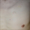
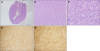
At the current admission, a skin examination revealed a 15×15 mm, firm, skin-colored, round-shaped nodule on his chest (Fig. 1). As skin metastases as well as other cystic diseases were suspected, an excisional biopsy was performed. The subsequent histopathologic examination revealed a well-demarcated subcutaneous tumor nodule. The tumor was mainly composed of atypical spindle and epithelioid type cells with various degrees of cytologic atypia. Multiple mitoses were also observed (Fig. 2A~C). Immunohistochemistry revealed positivity for c-kit (CD117), DOG-1 and negativity for smooth muscle actin, S-100, and CD34 (Fig. 2D, E). Additional molecular genetic analysis demonstrated a deletion mutation in exon 11 of the KIT gene. The histopathological features of the nodule were consistent with a GIST.
Following the biopsy, chest computed tomography was performed to look for any remnant soft tissue tumors. However, no abnormal lesion was observed within the excised site. After four weeks later, a brain magnetic resonance imaging of follow work-up revealed multiple metastases at the orbital left superior rectus muscle and occipital scalp. Under the diagnosis of imatinib-resistant GISTs, a second-line therapy with sunitinib was tried. After six weeks later, his chest nodule was completely improved. Further work-up at six month follow-up, computed tomography of head, chest and abdomen revealed decreased mass in left superior rectus muscle and no change of the cutaneous lesion, implicating the stable disease state.
DISCUSSION
GISTs are mesenchymal neoplasms which account for less than 1% of all gastrointestinal malignancies1. About 70 % of GISTs involve the stomach and otherwise the small intestine and esophagus, colon and rectum. Duodenal GISTs represent approximately 4% to 5% of all GISTs234. The histopathological features of GISTs include spindle cell (70%) or epithelioid predominance (20%) and otherwise mixed phenotypes5. Immunohistochemistry would be helpful for the diagnosis because GISTs show immunoreactivity for CD117 (95%), CD34 (70%) and DOG-1, which is a complementary stain of CD117124. GISTs are usually benign with a malignant transformation rate of 10~30%1. The median disease free survival has been estimated as 66.1 months for primary tumors and 31.6 months for recurrent or metastases cases respectively6. Duodenal GISTs are associated with a poorer recurrence-free survival outcome than nonduodenal GISTs3.
With regard to metastatic GISTs, DeMatteo et al.7 has previously reviewed 200 cases and reported that 61% of the metastases showed liver involvement, 20% had an intra-abdominal involvement and 6% had bone involvement. Since 2002, eight cases of a cutaneous metastasis from a GIST have been described and one report estimated these occurrences to represent 1% of advanced GISTs158910. In practice, these lesions can be clinically confused with primary mesenchymal tumors, a sarcomatoid carcinoma, or an epidermal cyst. In addition, the histologic features of a metastatic cutaneous GIST may be similar to those of metastases of synovial sarcoma, dedifferentiated liposarcomas, leiomyosarcomas and spindle cell tumors158. Metastatic GISTs are considered to be a late progressive manifestation8. Moreover, their presence may be indicative of multiple metastases as a cutaneous metastasis from a primary sarcoma is often accompanied by widespread systemic metastases58. In support of this possibility, asymptomatic rapidly growing cutaneous nodule of the current case drives further work-up and finally makes it found additional metastases.
To our knowledge, there has been no previously reported case of a cutaneous metastasis from a duodenal GIST. Although GISTs have a low rate of malignancy, cutaneous lesions may reflect multiple metastases from these tumors. Early detection can help to achieve a complete cytoreduction of the recurrent and metastatic tumor burden5, thereby improving the survival of the patients. A full skin examination of patients with a medical history of GISTs would be advisable to detect any visible clue to multiple metastases. Duodenal GISTs should not be an exception.
 XML Download