

 PDF
PDF ePub
ePub Citation
 Print
Print


INTRODUCTION
Acquired brachial cutaneous dyschromatosis (ABCD) is an acquired pigmentary disorder that presents as chronic, asymptomatic, gray-brown, geographic-shaped patches consisting of hyperpigmented macules mingled with hypopigmented lesions on the dorsal aspect of the forearms. It is usually bilateral and distally distributed. Most cases of ABCD have been reported in middle-aged postmenopausal women with Fitzpatrick skin types III~IV. Additionally, the majority of cases also had accompanying poikiloderma of Civatte at other body sites1. On histologic examination, the pigmented lesion of ABCD showed epidermal atrophy, increased basal layer pigmentation, solar elastosis and superficial telangiectasia1. However, in contrast to poikiloderma, there is no pigmentary incontinence2. Two hypotheses on the etiopathogenesis of ABCD have been suggested. The first hypothesis suggested the association between ABCD and hypertension or antihypertensive agents, specifically angiotensin converting enzyme inhibitors (ACEIs). The other hypothesis proposed that cumulative solar damage may cause ABCD23. We report a case of a male patient with ABCD who had no history of hypertension and ACEI medication, which does not support the former two hypotheses.
CASE REPORT
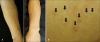
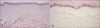
A 40-year-old Korean man presented to the dermatologic clinic with a complaint of multiple, reddish-brown colored macules on the outer aspects of both forearms (Fig. 1A). The patient did not remember when the lesions first appeared, but he stated that the discoloration had been present since at least the last four years, and it had spread gradually. He denied pruritus, pain or any other symptoms of skin lesions. He had neither an oral ulcer nor arthralgia. He did not have any specific medical or family history and his laboratory test results were in the normal range. Physical examination revealed mixed hyperpigmented and hypopigmented macules with focal atrophy and telangiectasia on both forearms (Fig. 1B). Punch biopsy was performed on the hyperpigmented macule on the outer side of his forearm. Histopathologic examination revealed epidermal atrophy and blunted rete ridges (Fig. 2A). Basal layer hyperpigmentation was remarkable which was highlighted with Fontana Masson stain for melanin (Fig. 2B). Several telangiectatic vessels were found in the upper dermis. There was no pigmentary incontinence. Scanty inflammatory cells were observed in the perivascular area, but solar elastosis was not remarkable. Congo red stain did not reveal amyloid deposit and periodic acid-Schiff stain did not show any fungal organism. Masson trichrome and elastic stain results were not remarkable. ABCD was diagnosed clinicopathologically and laser treatment was recommended. But, the patient refused treatment due to economic problems.
DISCUSSION
ABCD was first described in the report by of Rongioletti and Rebora1 who studied 20 Caucasian middle-aged patients from 1995 to 1998. In their study, the patient's age ranged from 46 to 72 years and all patients except one were women. In all patients, the lesions involved the dorsum of the forearms and presented as asymptomatic, irregular, gray-brown patches with geographic-shaped irregular borders, which were occasionally mixed with atrophic hypopigmented macules. The lesions were bilateral in all cases except one. According to the study by Rongioletti and Rebora1, 45 percent of patients (9 out of 20 patients) showed poikiloderma of Civatte on their neck and 65 percent of patients (13 out of 20 patients) had hypertension and were treated with anti-hypertensive drugs several years before the onset of pigmentation, especially 10 out of these 13 patients had been taking ACEIs. Histopathologic examination of the pigmented lesion of ABCD showed epidermal atrophy, hyperpigmentation in the basal layer, solar elastosis, and telangiectasia in the superficial dermis. Pigmentary incontinence and an increasing number of melanocytes were not observed3. Fontana Masson stain revealed that the pigment of brownish macules was melanin, and it was present along the basal cell layer with a homogeneous pattern.
Clinically, ABCD is similar to tinea versicolor, telangiectasia macularis eruptiva perstans (TMEP) and other disorders of pigmentation. Tinea versicolor can be easily ruled out by KOH-dissolved scale examination showing numerous budding yeasts and hyphae. TMEP shows a number of mast cells in the upper dermis, mainly around dilated vessels4. But our case showed no significant increase in mast cell number acquired bilateral telangiectatic macules (ABTM) is a newly described acquired pigmentary disorder5. ABTM shows irregular dark to brown telangiectatic macules on both upper arms, mostly in middle-aged men. But there are no hypopigmented atrophic macules in ABTM and our patient showed no upper arm skin lesions. Histologically, ABTM shows telangiectasia, capillary proliferation in the upper dermis, basal hyperpigmentation and melanogenic activity without mast cell infiltration. However, in contrast to our case, it shows no epidermal atrophy5. Lichen planus pigmentosus (LPP), a rare form of lichen planus, is characterized by mottled grey-brown macules on the sun-exposed areas such as face, neck, trunk, and limbs and also in the flexural folds. Interface dermatitis and melanophages are found in LPP on histologic examination, but they were not observed in our case6. Some drugs such as non-steroidal anti-inflammatory drugs, antimalarials, amiodarone, cytotoxic drugs, tetracycline, heavy metals and psychotropic drugs can cause pigmentary disorder78. An exhaustive medication history excluded drug-induced hyperpigmentation in our patient.
Currently, two hypotheses on the etiopathogenesis of ABCD have been proposed. The first hypothesis suggested the association of ABCD with hypertension and with antihypertensive medication, specifically ACEIs, and the other hypothesis proposed that ABCD is caused by cumulative sun damage. Based on the literature by Rongioletti and Rebora1, the authors observed that a large proportion of their cohort (13 out of 20 patients) had hypertension and had been treated with antihypertensive drugs. They suggested the association between ABCD and hypertension and antihypertensive medication, specifically ACEIs1. After the study by Rongioletti and Rebora1, there were two additional case reports in 20113 and 20142. On the other hand, Hu et al.3 and Abidi et al.2 reported the association with cumulative sun damage than with hypertension and ACEIs because most of the reported individuals had evidence of chronic sun exposure, such as poikiloderma of Civatte and solar elastosis. They also explained the association between ABCD and hypertension as an effect of high prevalence of hypertension23.
We reported a case of a middle-aged male patient with ABCD who had no history of hypertension and chronic sun exposure, and the presentation was distinct from that in the previously reported cases. Because ABCD has been described recently, further case reports are required to understand the clinical characteristics and pathogenesis of ABCD.
 XML Download