

 PDF
PDF ePub
ePub Citation
 Print
Print


Dear Editor:
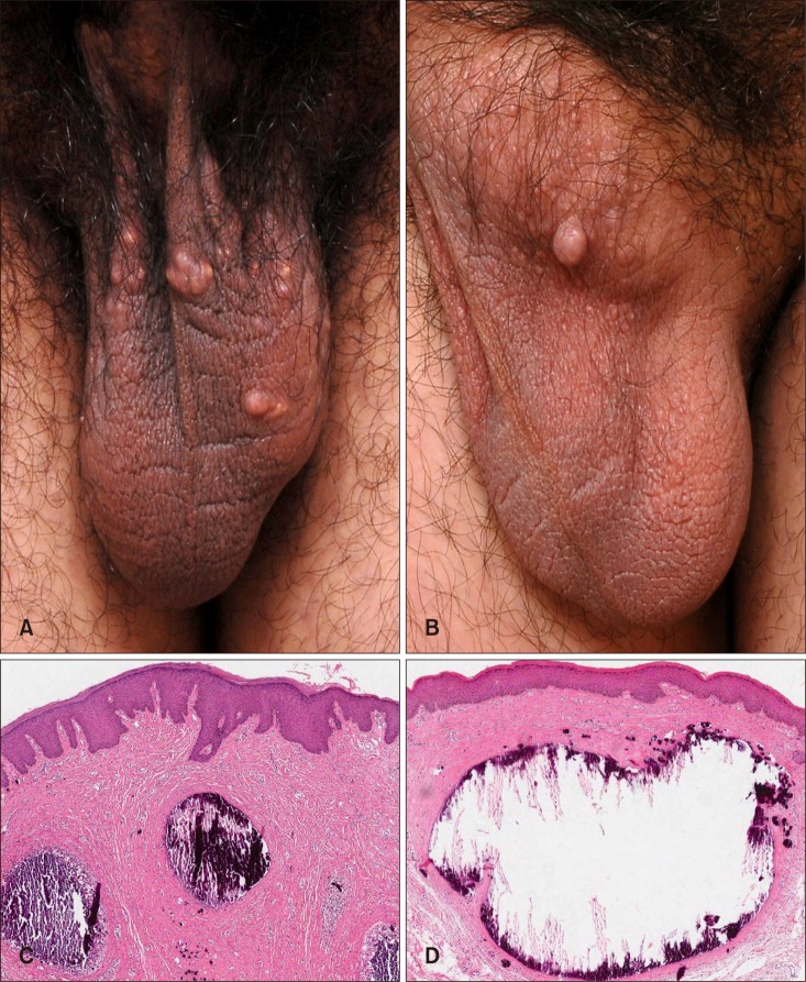
Scrotal calcinosis (SC) is a rare pathological condition characterized by painless, hard, asymptomatic nodules on scrotal skin without any tissue injury or metabolic derangement. The nodules are generally skin-colored, yellowish or white, and consist of calcium and phosphate deposits. SC usually affects patients in childhood or early adulthood. Because of the age of onset and location of occurrence, the condition may be a cause of embarrassment or misunderstanding. We describe the cases of two young adult patients with SC, who were brothers.
Two healthy Korean men, aged 22 and 23 years old, respectively, presented with scrotal nodules that gradually increased over time. The nodules first appeared in adolescence and increased in size and number during the previous 5 to 6 years. Both of the patients denied trauma, associated symptoms, or any prior treatments. There was no history of other systemic inflammatory or metabolic disease. Physical examination revealed multiple firm, non-tender, whitish papules on the scrotums of both patients (Fig. 1A, B). Skin biopsy was performed on both of the patients, and multiple calcium deposits and basophilic globules were found in the dermis (Fig. 1C, D). Routine laboratory examinations including serum calcium and phosphorus were all within normal limits. The possibility of tumoral calcinosis was ruled out, as both the patients had relatively small calcified nodules confined to the scrotum. The patients were diagnosed with SC and reassured of its benign nature; they remain in clinical follow-up.
SC was first described in 1883 by Leweinski and was established as a distinct entity by Shapiro et al. in 19701. It was initially termed idiopathic SC; however, although the etiology of the condition is still not fully elucidated, many physicians have come to believe that at least some SC is not genuinely idiopathic2. There are several major hypotheses for the origin. SC may be: 1) truly idiopathic, 2) secondary to calcification of epidermal inclusion cysts, 3) related to eccrine cysts of the scrotum, or 4) result from dystrophic calcification in the smooth muscle2. In our case, the calcifications were occasionally surrounded by histiocytes and giant cells, but did not have cystic membranes. However, we cannot rule out the possibility of epidermal cyst degeneration, despite the absence of epithelial elements3.
An extensive search of the literature did not find any familial cases of SC. Thus, we report the first case of SC in brothers. Multiple hypotheses have been considered in the pathogenesis of SC, but to be truly idiopathic, the possibility of patient factors (e.g., age, underlying morbidity, diet, medications, socioeconomic status, and genetic determinants) should be excluded. Our case suggests that SC, at least in part, may not be idiopathic4.
 XML Download