

 PDF
PDF ePub
ePub Citation
 Print
Print


Dear Editor:
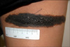
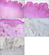
A 28-year-old woman at the 38th week of her first gestation presented with rapid enlarging papules and nodules in a 15 cm-sized black-pigmented plaque on her right posterior thigh. The plaque had been present since birth, and remained stable in size and pigmentation throughout her life. However, multiple clustered papules and nodules abruptly developed during the 2nd and 3rd trimester of her pregnancy (Fig. 1). To exclude the malignant melanoma arising within a congenital melanocytic nevus (CMN), a punch biopsy was performed. The histopathologic finding showed epidermal proliferation of melanocytes in the background of nevoid cells. The Ki-67 labeling index of the epidermal melanocytes mildly increased as 10% to 20%, whereas dermal melanocytes showed less than 5%. To exclude malignant melanoma, the patient underwent excision of the entire nevus. The distribution of pagetoid cells was mostly confined to the lower part of epidermis, without mitosis, necrosis and high-grade atypia (Fig. 2). The patient finally received the definite diagnosis of benign proliferative nodules (PNs) with mild atypia arising in
a CMN.
PNs usually represent the benign nodular proliferation of intradermal melanocytes1. PNs arising in a CMN occasionally need to be distinguished from melanoma, because of clinical characteristics such as rapid proliferation, hemorrhage, and ulceration. However, PNs have distinct histopathologic features: non-expanding, blending with the surrounding nevus, maturation, and benign prognosis2.
Interestingly, our patient presented abruptly, rapidly enlarging PNs during her pregnancy. According to previous studies, the leading opinion is that pregnancy cannot induce significant clinical and dermoscopical changes in melanocytic nevi, except for women with dysplastic nevus syndrome3. However, Chan et al.4 reported that histopathologic changes in melanocytic nevi during pregnancy had tendencies of higher mitotic rates, cellular atypia, and increased proliferation. Although these studies have focused on common nevi or dysplastic nevi rather than CMN, the results could partly explain the abrupt proliferation of atypical nevoid cells during pregnancy in our case.
The development of PNs in our patient might have been associated with hormonal changes during pregnancy. Estrogen receptor β (ER-β) expression is well known to be found in nevi and malignant melanomas, and to be influenced by hormonal changes in the pregnancy and post-partum periods3. Because ER-β suppresses the proliferation of nevoid cells and plays a protective role by inhibiting melanoma transformation, a decreased level of ER-β expression in CMN during pregnancy could be assumed to induce the development of PNs in CMN. However, There is not enough evidence of an association between ER-β expression level and histological atypia during pregnancy5.
In our case, although aggressive histological features including pagetoid spread of melanocytes and cellular atypia were mimicking malignant melanoma, the final diagnosis was benign PNs based on the overall benign architectural features. As shown by this case, the hormonal changes during pregnancy can be associated with various clinical and histopathological changes of CMN. However, even during pregnancy, whenever exclusion of malignant melanoma is required, proper procedures should be performed immediately.
 XML Download